 Movie
Movie Controller

[English] 日本語
 Yorodumi
- PDB-1g50: CRYSTAL STRUCTURE OF A WILD TYPE HER ALPHA LBD AT 2.9 ANGSTROM RE... -
+ Open data

- Basic information
| Entry | Database: PDB / ID: 1g50 | ||||||
|---|---|---|---|---|---|---|---|
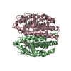
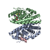
| Title | CRYSTAL STRUCTURE OF A WILD TYPE HER ALPHA LBD AT 2.9 ANGSTROM RESOLUTION | ||||||
 Components Components | ESTROGEN RECEPTOR | ||||||
 Keywords Keywords | DNA BINDING PROTEIN / alpha helices / estradiol / Structural Proteomics in Europe / SPINE / Structural Genomics | ||||||
| Function / homology |  Function and homology information Function and homology informationregulation of epithelial cell apoptotic process / steroid hormone receptor signaling pathway / antral ovarian follicle growth / regulation of branching involved in prostate gland morphogenesis / RUNX1 regulates transcription of genes involved in WNT signaling / RUNX1 regulates estrogen receptor mediated transcription / nuclear estrogen receptor activity / regulation of toll-like receptor signaling pathway / prostate epithelial cord elongation / epithelial cell proliferation involved in mammary gland duct elongation ...regulation of epithelial cell apoptotic process / steroid hormone receptor signaling pathway / antral ovarian follicle growth / regulation of branching involved in prostate gland morphogenesis / RUNX1 regulates transcription of genes involved in WNT signaling / RUNX1 regulates estrogen receptor mediated transcription / nuclear estrogen receptor activity / regulation of toll-like receptor signaling pathway / prostate epithelial cord elongation / epithelial cell proliferation involved in mammary gland duct elongation / prostate epithelial cord arborization involved in prostate glandular acinus morphogenesis / epithelial cell development / mammary gland branching involved in pregnancy / uterus development / vagina development / TFIIB-class transcription factor binding / androgen metabolic process / negative regulation of smooth muscle cell apoptotic process / nuclear receptor-mediated steroid hormone signaling pathway / mammary gland alveolus development / : / cellular response to estrogen stimulus / estrogen response element binding / : / Mitochondrial unfolded protein response (UPRmt) / Nuclear signaling by ERBB4 / RNA polymerase II preinitiation complex assembly / positive regulation of nitric-oxide synthase activity / estrogen receptor signaling pathway / steroid binding / TFAP2 (AP-2) family regulates transcription of growth factors and their receptors / protein localization to chromatin / 14-3-3 protein binding / ESR-mediated signaling / negative regulation of miRNA transcription / TBP-class protein binding / nitric-oxide synthase regulator activity / nuclear estrogen receptor binding / transcription corepressor binding / transcription coregulator binding / stem cell differentiation / negative regulation of canonical NF-kappaB signal transduction / SUMOylation of intracellular receptors / cellular response to estradiol stimulus / euchromatin / Nuclear Receptor transcription pathway / response to estrogen / beta-catenin binding / male gonad development / nuclear receptor activity / positive regulation of fibroblast proliferation / transcription coactivator binding / Constitutive Signaling by Aberrant PI3K in Cancer / positive regulation of nitric oxide biosynthetic process / sequence-specific double-stranded DNA binding / Regulation of RUNX2 expression and activity / Ovarian tumor domain proteases / response to estradiol / PIP3 activates AKT signaling / ATPase binding / PI5P, PP2A and IER3 Regulate PI3K/AKT Signaling / positive regulation of cytosolic calcium ion concentration / regulation of inflammatory response / fibroblast proliferation / DNA-binding transcription activator activity, RNA polymerase II-specific / transcription regulator complex / Estrogen-dependent gene expression / phospholipase C-activating G protein-coupled receptor signaling pathway / DNA-binding transcription factor activity, RNA polymerase II-specific / calmodulin binding / Extra-nuclear estrogen signaling / RNA polymerase II cis-regulatory region sequence-specific DNA binding / chromatin remodeling / DNA-binding transcription factor activity / negative regulation of gene expression / chromatin binding / regulation of transcription by RNA polymerase II / regulation of DNA-templated transcription / protein kinase binding / positive regulation of DNA-templated transcription / chromatin / enzyme binding / negative regulation of transcription by RNA polymerase II / Golgi apparatus / signal transduction / positive regulation of transcription by RNA polymerase II / protein-containing complex / zinc ion binding / nucleoplasm / membrane / identical protein binding / nucleus / plasma membrane / cytoplasm / cytosol Similarity search - Function | ||||||
| Biological species |  Homo sapiens (human) Homo sapiens (human) | ||||||
| Method |  X-RAY DIFFRACTION / SYNCHROTRON / Resolution: 2.9 Å X-RAY DIFFRACTION / SYNCHROTRON / Resolution: 2.9 Å | ||||||
 Authors Authors | Eiler, S. / Gangloff, M. / Duclaud, S. / Moras, D. / Ruff, M. / Structural Proteomics in Europe (SPINE) | ||||||
 Citation Citation | Journal: Protein Expr.Purif. / Year: 2001 Title: Overexpression, Purification, and Crystal Structure of Native ER alpha LBD Authors: Eiler, S. / Gangloff, M. / Duclaud, S. / Moras, D. / Ruff, M. | ||||||
| History |
|
- Structure visualization
| Structure viewer | Molecule: MolmilJmol/JSmol |
|---|
- Downloads & links
-Download
| PDBx/mmCIF format | 1g50.cif.gz | 160.8 KB | Display | PDBx/mmCIF format |
|---|---|---|---|---|
| PDB format | pdb1g50.ent.gz | 126.7 KB | Display | PDB format |
| PDBx/mmJSON format | 1g50.json.gz | Tree view | PDBx/mmJSON format | |
| Others |  Other downloads Other downloads |
-Validation report
| Arichive directory | https://data.pdbj.org/pub/pdb/validation_reports/g5/1g50ftp://data.pdbj.org/pub/pdb/validation_reports/g5/1g50 | HTTPS FTP |
|---|
-Related structure data
| Related structure data | 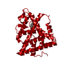 1qktS S: Starting model for refinement |
|---|---|
| Similar structure data | |
| Other databases |


-Links
 PDBj
PDBj


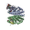
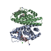
- Assembly
| Deposited unit | 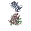
| ||||||||
|---|---|---|---|---|---|---|---|---|---|
| 1 | 
| ||||||||
| 2 | 
| ||||||||
| Unit cell |
|
-Components
| #1: Protein | Mass: 28274.289 Da / Num. of mol.: 3 Source method: isolated from a genetically manipulated source Source: (gene. exp.) Homo sapiens (human) / Plasmid: PET15B / Species (production host): Escherichia coli / Production host:  #2: Chemical |   Mass: 272.382 Da / Num. of mol.: 3 / Source method: obtained synthetically / Formula: C18H24O2 Mass: 272.382 Da / Num. of mol.: 3 / Source method: obtained synthetically / Formula: C18H24O2#3: Water | ChemComp-HOH / |  Mass: 18.015 Da / Num. of mol.: 165 / Source method: isolated from a natural source / Formula: H2O Mass: 18.015 Da / Num. of mol.: 165 / Source method: isolated from a natural source / Formula: H2O |
|---|
-Experimental details
-Experiment
| Experiment | Method: X-RAY DIFFRACTION / Number of used crystals: 1 |
|---|
- Sample preparation
| Crystal | Density Matthews: 2.58 Å3/Da / Density % sol: 52.26 % | ||||||||||||||||||||||||||||||||||||||||||||||||||||||||||||||||||||||||||||||||||||||||||||||||||
|---|---|---|---|---|---|---|---|---|---|---|---|---|---|---|---|---|---|---|---|---|---|---|---|---|---|---|---|---|---|---|---|---|---|---|---|---|---|---|---|---|---|---|---|---|---|---|---|---|---|---|---|---|---|---|---|---|---|---|---|---|---|---|---|---|---|---|---|---|---|---|---|---|---|---|---|---|---|---|---|---|---|---|---|---|---|---|---|---|---|---|---|---|---|---|---|---|---|---|---|
| Crystal grow | Temperature: 277 K / Method: vapor diffusion, hanging drop / pH: 7.1 Details: PEG400, pH 7.1, VAPOR DIFFUSION, HANGING DROP, temperature 277K | ||||||||||||||||||||||||||||||||||||||||||||||||||||||||||||||||||||||||||||||||||||||||||||||||||
| Crystal grow | *PLUS Temperature: 4 ℃ / pH: 7.3 | ||||||||||||||||||||||||||||||||||||||||||||||||||||||||||||||||||||||||||||||||||||||||||||||||||
| Components of the solutions | *PLUS
|
-Data collection
| Diffraction | Mean temperature: 110 K |
|---|---|
| Diffraction source | Source: SYNCHROTRON / Site: ESRF  / Beamline: BM30A / Wavelength: 0.9934 Å / Beamline: BM30A / Wavelength: 0.9934 Å |
| Detector | Detector: CCD / Date: Apr 1, 1997 |
| Radiation | Protocol: SINGLE WAVELENGTH / Monochromatic (M) / Laue (L): M / Scattering type: x-ray |
| Radiation wavelength | Wavelength: 0.9934 Å / Relative weight: 1 |
| Reflection | Resolution: 2.9→40 Å / Num. all: 20000 / Num. obs: 18142 / % possible obs: 91 % / Observed criterion σ(F): 0 / Observed criterion σ(I): 0 / Redundancy: 1.8 % / Biso Wilson estimate: 34.9 Å2 / Rsym value: 8 |
| Reflection | *PLUS % possible obs: 91 % / Rmerge(I) obs: 0.08 |
| Reflection shell | *PLUS Highest resolution: 2.9 Å / Lowest resolution: 3.1 Å / % possible obs: 78 % |
- Processing
| Software |
| ||||||||||||||||||||||||||||||||||||||||
|---|---|---|---|---|---|---|---|---|---|---|---|---|---|---|---|---|---|---|---|---|---|---|---|---|---|---|---|---|---|---|---|---|---|---|---|---|---|---|---|---|---|
| Refinement | Starting model: pdb entry 1qkt Resolution: 2.9→49.18 Å / Rfactor Rfree error: 0.007 / Data cutoff high absF: 1999535.89 / Data cutoff high rms absF: 1999535.89 / Data cutoff low absF: 0 / Isotropic thermal model: RESTRAINED / Cross valid method: THROUGHOUT / σ(F): 0
| ||||||||||||||||||||||||||||||||||||||||
| Solvent computation | Solvent model: FLAT MODEL / Bsol: 44.13 Å2 / ksol: 0.322 e/Å3 | ||||||||||||||||||||||||||||||||||||||||
| Displacement parameters | Biso mean: 61.2 Å2
| ||||||||||||||||||||||||||||||||||||||||
| Refine analyze |
| ||||||||||||||||||||||||||||||||||||||||
| Refinement step | Cycle: LAST / Resolution: 2.9→49.18 Å
| ||||||||||||||||||||||||||||||||||||||||
| Refine LS restraints |
| ||||||||||||||||||||||||||||||||||||||||
| LS refinement shell | Resolution: 2.9→3.08 Å / Rfactor Rfree error: 0.029 / Total num. of bins used: 6
| ||||||||||||||||||||||||||||||||||||||||
| Xplor file |
| ||||||||||||||||||||||||||||||||||||||||
| Software | *PLUS Name: CNS / Classification: refinement | ||||||||||||||||||||||||||||||||||||||||
| Refinement | *PLUS Lowest resolution: 15 Å / σ(F): 0 / % reflection Rfree: 9.8 % / Rfactor Rfree: 0.31 | ||||||||||||||||||||||||||||||||||||||||
| Solvent computation | *PLUS | ||||||||||||||||||||||||||||||||||||||||
| Displacement parameters | *PLUS Biso mean: 61.2 Å2 | ||||||||||||||||||||||||||||||||||||||||
| Refine LS restraints | *PLUS
| ||||||||||||||||||||||||||||||||||||||||
| LS refinement shell | *PLUS Rfactor Rfree: 0.465 / % reflection Rfree: 9.9 % / Rfactor Rwork: 0.382 |
