 Movie
Movie Controller


+ Open data

- Basic information
| Entry | Database: PDB / ID: 1g3g | |||||||||
|---|---|---|---|---|---|---|---|---|---|---|
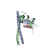
| Title | NMR STRUCTURE OF THE FHA1 DOMAIN OF YEAST RAD53 | |||||||||
 Components Components | PROTEIN KINASE SPK1 | |||||||||
 Keywords Keywords | TRANSFERASE / FHA domain / Rad53 / Phosphopeptide / Phosphoprotein | |||||||||
| Function / homology |  Function and homology information Function and homology informationG2/M DNA damage checkpoint / Chk1/Chk2(Cds1) mediated inactivation of Cyclin B:Cdk1 complex / deoxyribonucleoside triphosphate biosynthetic process / mitotic DNA damage checkpoint signaling / negative regulation of phosphorylation / dual-specificity kinase / Recruitment and ATM-mediated phosphorylation of repair and signaling proteins at DNA double strand breaks / Ubiquitin-Mediated Degradation of Phosphorylated Cdc25A / telomere maintenance in response to DNA damage / negative regulation of DNA damage checkpoint ...G2/M DNA damage checkpoint / Chk1/Chk2(Cds1) mediated inactivation of Cyclin B:Cdk1 complex / deoxyribonucleoside triphosphate biosynthetic process / mitotic DNA damage checkpoint signaling / negative regulation of phosphorylation / dual-specificity kinase / Recruitment and ATM-mediated phosphorylation of repair and signaling proteins at DNA double strand breaks / Ubiquitin-Mediated Degradation of Phosphorylated Cdc25A / telomere maintenance in response to DNA damage / negative regulation of DNA damage checkpoint / DNA replication origin binding / DNA replication initiation / regulation of DNA repair / protein serine/threonine/tyrosine kinase activity / DNA damage checkpoint signaling / intracellular protein localization / protein tyrosine kinase activity / protein kinase activity / protein serine kinase activity / DNA repair / protein serine/threonine kinase activity / ATP binding / nucleus / cytosol / cytoplasm Similarity search - Function | |||||||||
| Biological species |  | |||||||||
| Method | SOLUTION NMR / simulated annealing | |||||||||
 Authors Authors | Yuan, C. / Liao, H. / Su, M. / Yongkiettrakul, S. / Byeon, I.-J.L. / Tsai, M.-D. | |||||||||
 Citation Citation | Journal: J.Mol.Biol. / Year: 2000 Title: Structure of the FHA1 domain of yeast Rad53 and identification of binding sites for both FHA1 and its target protein Rad9 Authors: Liao, H. / Yuan, C. / Su, M.I. / Yongkiettrakul, S. / Qin, D. / Li, H. / Byeon, I.J. / Pei, D. / Tsai, M.D. | |||||||||
| History |
|
- Structure visualization
| Structure viewer | Molecule: MolmilJmol/JSmol |
|---|
- Downloads & links
-Download
| PDBx/mmCIF format | 1g3g.cif.gz | 1019.6 KB | Display | PDBx/mmCIF format |
|---|---|---|---|---|
| PDB format | pdb1g3g.ent.gz | 850.7 KB | Display | PDB format |
| PDBx/mmJSON format | 1g3g.json.gz | Tree view | PDBx/mmJSON format | |
| Others |  Other downloads Other downloads |
-Validation report
| Arichive directory | https://data.pdbj.org/pub/pdb/validation_reports/g3/1g3gftp://data.pdbj.org/pub/pdb/validation_reports/g3/1g3g | HTTPS FTP |
|---|
-Related structure data
| Similar structure data |
|---|


-Links
 PDBj
PDBj


- Assembly
| Deposited unit | 
| |||||||||
|---|---|---|---|---|---|---|---|---|---|---|
| 1 |
| |||||||||
| NMR ensembles |
|
-Components
| #1: Protein | Mass: 18506.938 Da / Num. of mol.: 1 / Fragment: THE N-TERMINAL FHA DOMAIN (FHA1), RESIDUES 1-164 / Mutation: M1G Source method: isolated from a genetically manipulated source Source: (gene. exp.) Gene: SPK1 OR RAD53 / Plasmid: PGEX-4T / Species (production host): Escherichia coli / Production host:  References: UniProt: P22216, Transferases; Transferring phosphorus-containing groups; Phosphotransferases with an alcohol group as acceptor |
|---|
-Experimental details
-Experiment
| Experiment | Method: SOLUTION NMR | ||||||||||||
|---|---|---|---|---|---|---|---|---|---|---|---|---|---|
| NMR experiment |
| ||||||||||||
| NMR details | Text: The structure was determined using triple-resonance NMR spectroscopy. |
- Sample preparation
| Details | Contents: 0.5 mM protein U-15N, 13C; 10 mM sodium phosphate buffer (pH 6.5), 1 mM DTT, and 1 mM EDTA; 95 % H2O, 5 % D2O Solvent system: 95% H2O/5% D2O |
|---|---|
| Sample conditions | Ionic strength: 10 mM sodium phosphate, 1 mM DTT, and 1 mM EDTA pH: 6.5 / Pressure: ambient / Temperature: 293 K |
| Crystal grow | *PLUS Method: other / Details: NMR |
-NMR measurement
| NMR spectrometer | Type: Bruker AVANCE / Manufacturer: Bruker / Model: AVANCE / Field strength: 800 MHz |
|---|
- Processing
| NMR software |
| ||||||||||||||||||||
|---|---|---|---|---|---|---|---|---|---|---|---|---|---|---|---|---|---|---|---|---|---|
| Refinement | Method: simulated annealing / Software ordinal: 1 Details: The structures are based on a total of 2156 restraints, 1886 are NOE-derived distance constraints, 192 TALOS-derived dihedral angle restraints,78 distance restraints from hydrogen bonds. ...Details: The structures are based on a total of 2156 restraints, 1886 are NOE-derived distance constraints, 192 TALOS-derived dihedral angle restraints,78 distance restraints from hydrogen bonds. RESIDUES 1-14 ARE POORLY DEFINED BY THE EXPERIMENTAL DATA. THUS, NO MEANING SHOULD BE GIVEN TO THOSE RESIDUES' COORDINATES. | ||||||||||||||||||||
| NMR representative | Selection criteria: closest to the average | ||||||||||||||||||||
| NMR ensemble | Conformer selection criteria: structures with the least restraint violations,structures with the lowest energy Conformers calculated total number: 50 / Conformers submitted total number: 20 |
 X-PLOR