 Movie
Movie Controller

[English] 日本語
 Yorodumi
- PDB-1fu5: NMR STRUCTURE OF THE N-SH2 DOMAIN OF THE P85 SUBUNIT OF PI3-KINAS... -
+ Open data

- Basic information
| Entry | Database: PDB / ID: 1fu5 | ||||||
|---|---|---|---|---|---|---|---|
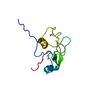
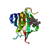
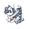
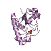
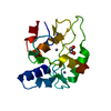
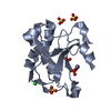
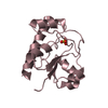
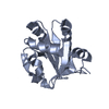
| Title | NMR STRUCTURE OF THE N-SH2 DOMAIN OF THE P85 SUBUNIT OF PI3-KINASE COMPLEXED TO A DOUBLY PHOSPHORYLATED PEPTIDE DERIVED FROM POLYOMAVIRUS MIDDLE T ANTIGEN | ||||||
 Components Components |
| ||||||
 Keywords Keywords | PEPTIDE BINDING PROTEIN / protein-peptide complex | ||||||
| Function / homology |  Function and homology information Function and homology informationPI3K events in ERBB4 signaling / CD28 dependent PI3K/Akt signaling / MET activates PI3K/AKT signaling / Erythropoietin activates Phosphoinositide-3-kinase (PI3K) / Interleukin receptor SHC signaling / Co-stimulation by ICOS / Tie2 Signaling / Interleukin-3, Interleukin-5 and GM-CSF signaling / FLT3 Signaling / GAB1 signalosome ...PI3K events in ERBB4 signaling / CD28 dependent PI3K/Akt signaling / MET activates PI3K/AKT signaling / Erythropoietin activates Phosphoinositide-3-kinase (PI3K) / Interleukin receptor SHC signaling / Co-stimulation by ICOS / Tie2 Signaling / Interleukin-3, Interleukin-5 and GM-CSF signaling / FLT3 Signaling / GAB1 signalosome / PI3K events in ERBB2 signaling / PI-3K cascade:FGFR1 / PI-3K cascade:FGFR3 / PI-3K cascade:FGFR4 / RHOJ GTPase cycle / IRS-mediated signalling / Interleukin-7 signaling / GP1b-IX-V activation signalling / Signaling by LTK / Synthesis of PIPs at the plasma membrane / PI3K/AKT activation / PI-3K cascade:FGFR2 / Regulation of signaling by CBL / RND1 GTPase cycle / RND2 GTPase cycle / Downstream TCR signaling / RHOF GTPase cycle / PI3K Cascade / Signaling by SCF-KIT / Role of LAT2/NTAL/LAB on calcium mobilization / VEGFA-VEGFR2 Pathway / RHOB GTPase cycle / RND3 GTPase cycle / CDC42 GTPase cycle / RAC3 GTPase cycle / GPVI-mediated activation cascade / RHOC GTPase cycle / PIP3 activates AKT signaling / Downstream signal transduction / Signaling by ALK / Role of phospholipids in phagocytosis / RHOU GTPase cycle / RHOV GTPase cycle / Antigen activates B Cell Receptor (BCR) leading to generation of second messengers / RAC2 GTPase cycle / DAP12 signaling / RHOG GTPase cycle / RET signaling / RAC1 GTPase cycle / RHOA GTPase cycle / negative regulation of muscle cell apoptotic process / Extra-nuclear estrogen signaling / PI5P, PP2A and IER3 Regulate PI3K/AKT Signaling / perinuclear endoplasmic reticulum membrane / regulation of toll-like receptor 4 signaling pathway / phosphatidylinositol metabolic process / response to yeast / phosphatidylinositol 3-kinase regulator activity / 1-phosphatidylinositol-3-kinase regulator activity / positive regulation of endoplasmic reticulum unfolded protein response / T follicular helper cell differentiation / response to fatty acid / interleukin-18-mediated signaling pathway / response to fructose / phosphatidylinositol 3-kinase regulatory subunit binding / response to growth factor / neurotrophin TRKA receptor binding / positive regulation of focal adhesion disassembly / negative regulation of cell adhesion / cis-Golgi network / ErbB-3 class receptor binding / negative regulation of stress fiber assembly / phosphatidylinositol 3-kinase complex / regulation of stress fiber assembly / cellular response to fatty acid / platelet-derived growth factor receptor binding / positive regulation of synapse assembly / negative regulation of cell-cell adhesion / kinase activator activity / RAF/MAP kinase cascade / G alpha (q) signalling events / phosphatidylinositol 3-kinase complex, class IA / positive regulation of filopodium assembly / growth hormone receptor signaling pathway / insulin binding / natural killer cell mediated cytotoxicity / negative regulation of heart rate / negative regulation of osteoclast differentiation / 1-phosphatidylinositol-3-kinase activity / response to dexamethasone / response to testosterone / response to iron(II) ion / phosphatidylinositol phosphate biosynthetic process / negative regulation of cell-matrix adhesion / extrinsic apoptotic signaling pathway via death domain receptors / viral process / insulin receptor substrate binding / response to amino acid / membrane => GO:0016020 / host cell membrane Similarity search - Function | ||||||
| Biological species |  | ||||||
| Method | SOLUTION NMR / The structures were energy minimized with MSI DISCOVER. | ||||||
 Authors Authors | Weber, T. / Schaffhausen, B. / Liu, Y. / Guenther, U.L. | ||||||
 Citation Citation | Journal: Biochemistry / Year: 2000 Title: NMR structure of the N-SH2 of the p85 subunit of phosphoinositide 3-kinase complexed to a doubly phosphorylated peptide reveals a second phosphotyrosine binding site. Authors: Weber, T. / Schaffhausen, B. / Liu, Y. / Gunther, U.L. | ||||||
| History |
|
- Structure visualization
| Structure viewer | Molecule: MolmilJmol/JSmol |
|---|
- Downloads & links
-Download
| PDBx/mmCIF format | 1fu5.cif.gz | 53.6 KB | Display | PDBx/mmCIF format |
|---|---|---|---|---|
| PDB format | pdb1fu5.ent.gz | 39 KB | Display | PDB format |
| PDBx/mmJSON format | 1fu5.json.gz | Tree view | PDBx/mmJSON format | |
| Others |  Other downloads Other downloads |
-Validation report
| Arichive directory | https://data.pdbj.org/pub/pdb/validation_reports/fu/1fu5ftp://data.pdbj.org/pub/pdb/validation_reports/fu/1fu5 | HTTPS FTP |
|---|
-Related structure data


-Links
 PDBj
PDBj


- Assembly
| Deposited unit | 
| |||||||||
|---|---|---|---|---|---|---|---|---|---|---|
| 1 |
| |||||||||
| NMR ensembles |
|
-Components
| #1: Protein | Mass: 12870.384 Da / Num. of mol.: 1 Fragment: RESIDUES 321 TO 431 OF P85, N-SH2 (SRC HOMOLOGY 2) DOMAIN Source method: isolated from a genetically manipulated source Source: (gene. exp.)  |
|---|---|
| #2: Protein/peptide | Mass: 2063.089 Da / Num. of mol.: 1 Fragment: RESIDUES 312 TO 326 OF MT ANTIGEN, Y315 AND Y322 PHOSPHORYLATED Source method: obtained synthetically Details: MT peptide was synthesized by the Tufts Protein Chemistry Facility References: UniProt: P03076, UniProt: P03077*PLUS |
| Has protein modification | Y |
-Experimental details
-Experiment
| Experiment | Method: SOLUTION NMR | ||||||||||||||||||||
|---|---|---|---|---|---|---|---|---|---|---|---|---|---|---|---|---|---|---|---|---|---|
| NMR experiment |
| ||||||||||||||||||||
| NMR details | Text: NOESY assignments were obtained by a semi-automatic procedure employing a program from Pristovsek [Pristovsek, P. & Kidric, J. (1997) Biopol. 42, 671-679)]. Initial calculations included only ...Text: NOESY assignments were obtained by a semi-automatic procedure employing a program from Pristovsek [Pristovsek, P. & Kidric, J. (1997) Biopol. 42, 671-679)]. Initial calculations included only intramolecular constraints. The observed NOEs derived from 13C{F1}-filtered 2D-NOESY spectra were incorporated into the structure calculation when the protein fold was already correct. |
- Sample preparation
| Details | Contents: 0.15mM N-SH2 15N, 13C; MT peptide; 0.1mM KCl; 95% H2O, 5% D2O Solvent system: 100% H2O |
|---|---|
| Sample conditions | Ionic strength: 0.1mM / pH: 6.8 / Pressure: 1 bar / Temperature: 305 K |
| Crystal grow | *PLUS Method: other / Details: NMR |
-NMR measurement
| NMR spectrometer |
|
|---|
- Processing
| NMR software |
| ||||||||||||||||||
|---|---|---|---|---|---|---|---|---|---|---|---|---|---|---|---|---|---|---|---|
| Refinement | Method: The structures were energy minimized with MSI DISCOVER. Software ordinal: 1 / Details: The structure with the lowest energy is presented | ||||||||||||||||||
| NMR representative | Selection criteria: lowest energy | ||||||||||||||||||
| NMR ensemble | Conformer selection criteria: structures with favorable non-bond energy Conformers calculated total number: 110 / Conformers submitted total number: 1 |
