 ムービー
ムービー コントローラー


+ データを開く

- 基本情報
基本情報
| 登録情報 | データベース: PDB / ID: 1ftk | ||||||
|---|---|---|---|---|---|---|---|
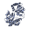
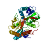
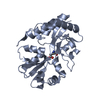
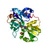
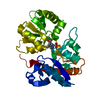
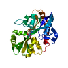
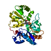
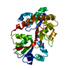
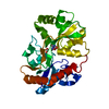
| タイトル | CRYSTAL STRUCTURE OF THE GLUR2 LIGAND BINDING CORE (S1S2I) IN COMPLEX WITH KAINATE AT 1.6 A RESOLUTION | ||||||
 要素 要素 | GLUTAMATE RECEPTOR SUBUNIT 2 | ||||||
 キーワード キーワード | MEMBRANE PROTEIN / GluR2 / S1S2 / ligand binding domain / kainate / partial agonist / ionotropic glutamate receptor | ||||||
| 機能・相同性 |  機能・相同性情報 機能・相同性情報spine synapse / dendritic spine neck / dendritic spine head / cellular response to amine stimulus / perisynaptic space / Activation of AMPA receptors / ligand-gated monoatomic cation channel activity / AMPA glutamate receptor activity / response to lithium ion / Trafficking of GluR2-containing AMPA receptors ...spine synapse / dendritic spine neck / dendritic spine head / cellular response to amine stimulus / perisynaptic space / Activation of AMPA receptors / ligand-gated monoatomic cation channel activity / AMPA glutamate receptor activity / response to lithium ion / Trafficking of GluR2-containing AMPA receptors / kainate selective glutamate receptor activity / AMPA glutamate receptor complex / cellular response to glycine / extracellularly glutamate-gated ion channel activity / immunoglobulin binding / asymmetric synapse / ionotropic glutamate receptor complex / conditioned place preference / regulation of receptor recycling / glutamate receptor binding / Unblocking of NMDA receptors, glutamate binding and activation / positive regulation of synaptic transmission / regulation of synaptic transmission, glutamatergic / response to fungicide / cytoskeletal protein binding / glutamate-gated receptor activity / regulation of long-term synaptic depression / extracellular ligand-gated monoatomic ion channel activity / cellular response to brain-derived neurotrophic factor stimulus / presynaptic active zone membrane / glutamate-gated calcium ion channel activity / somatodendritic compartment / dendrite membrane / ionotropic glutamate receptor binding / ligand-gated monoatomic ion channel activity involved in regulation of presynaptic membrane potential / ionotropic glutamate receptor signaling pathway / dendrite cytoplasm / synaptic membrane / dendritic shaft / SNARE binding / transmitter-gated monoatomic ion channel activity involved in regulation of postsynaptic membrane potential / synaptic transmission, glutamatergic / PDZ domain binding / protein tetramerization / establishment of protein localization / postsynaptic density membrane / cerebral cortex development / modulation of chemical synaptic transmission / receptor internalization / Schaffer collateral - CA1 synapse / terminal bouton / synaptic vesicle / synaptic vesicle membrane / presynapse / signaling receptor activity / amyloid-beta binding / presynaptic membrane / growth cone / scaffold protein binding / perikaryon / chemical synaptic transmission / dendritic spine / postsynaptic membrane / neuron projection / postsynaptic density / axon / external side of plasma membrane / neuronal cell body / dendrite / synapse / protein kinase binding / protein-containing complex binding / glutamatergic synapse / cell surface / endoplasmic reticulum / protein-containing complex / identical protein binding / membrane / plasma membrane 類似検索 - 分子機能 | ||||||
| 生物種 |  | ||||||
| 手法 |  X線回折 / シンクロトロン / 解像度: 1.6 Å X線回折 / シンクロトロン / 解像度: 1.6 Å | ||||||
 データ登録者 データ登録者 | Gouaux, E. / Armstrong, N. | ||||||
 引用 引用 | ジャーナル: Neuron / 年: 2000 タイトル: Mechanisms for activation and antagonism of an AMPA-sensitive glutamate receptor: crystal structures of the GluR2 ligand binding core. 著者: Armstrong, N. / Gouaux, E. #1: ジャーナル: Protein Sci. / 年: 1998タイトル: Probing the ligand binding domain of the GluR2 receptor by proteolysis and deletion mutagenesis defines domain boundaries and yields a crystallizable construct 著者: Chen, G.Q. / Sun, Y. / Jin, R. / Gouaux, E. | ||||||
| 履歴 |
|
- 構造の表示
構造の表示
| 構造ビューア | 分子: MolmilJmol/JSmol |
|---|
- ダウンロードとリンク
-ダウンロード
| PDBx/mmCIF形式 | 1ftk.cif.gz | 66.8 KB | 表示 | PDBx/mmCIF形式 |
|---|---|---|---|---|
| PDB形式 | pdb1ftk.ent.gz | 47.5 KB | 表示 | PDB形式 |
| PDBx/mmJSON形式 | 1ftk.json.gz | ツリー表示 | PDBx/mmJSON形式 | |
| その他 |  その他のダウンロード その他のダウンロード |
-検証レポート
| 文書・要旨 | 1ftk_validation.pdf.gz | 385.2 KB | 表示 | wwPDB検証レポート |
|---|---|---|---|---|
| 文書・詳細版 | 1ftk_full_validation.pdf.gz | 386.7 KB | 表示 | |
| XML形式データ | 1ftk_validation.xml.gz | 6.4 KB | 表示 | |
| CIF形式データ | 1ftk_validation.cif.gz | 10.8 KB | 表示 | |
| アーカイブディレクトリ | https://data.pdbj.org/pub/pdb/validation_reports/ft/1ftkftp://data.pdbj.org/pub/pdb/validation_reports/ft/1ftk | HTTPS FTP |
-関連構造データ


-リンク
 PDBj
PDBj


- 集合体
集合体
| 登録構造単位 | 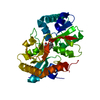
| ||||||||
|---|---|---|---|---|---|---|---|---|---|
| 1 |
| ||||||||
| 単位格子 |
|
-要素
| #1: タンパク質 | 分子量: 30741.197 Da / 分子数: 1 / 断片: LIGAND BINDING CORE, S1S2I / 由来タイプ: 組換発現 / 由来: (組換発現)  |
|---|---|
| #2: 化合物 | ChemComp-KAI /   分子量: 213.230 Da / 分子数: 1 / 由来タイプ: 合成 / 式: C10H15NO4 分子量: 213.230 Da / 分子数: 1 / 由来タイプ: 合成 / 式: C10H15NO4 |
| #3: 水 | ChemComp-HOH /  分子量: 18.015 Da / 分子数: 250 / 由来タイプ: 天然 / 式: H2O 分子量: 18.015 Da / 分子数: 250 / 由来タイプ: 天然 / 式: H2O |
| Has protein modification | Y |
-実験情報
-実験
| 実験 | 手法: X線回折 / 使用した結晶の数: 1 |
|---|
- 試料調製
試料調製
| 結晶 | マシュー密度: 2.06 Å3/Da / 溶媒含有率: 40.34 % | ||||||||||||||||||||||||||||||||||||||||||
|---|---|---|---|---|---|---|---|---|---|---|---|---|---|---|---|---|---|---|---|---|---|---|---|---|---|---|---|---|---|---|---|---|---|---|---|---|---|---|---|---|---|---|---|
| 結晶化 | 温度: 277 K / 手法: 蒸気拡散法, ハンギングドロップ法 / pH: 6.5 詳細: 15% PEG 8000 50 mM potassium phosphate , pH 6.5, VAPOR DIFFUSION, HANGING DROP, temperature 277K | ||||||||||||||||||||||||||||||||||||||||||
| 結晶化 | *PLUS 温度: 4 ℃ / pH: 7 | ||||||||||||||||||||||||||||||||||||||||||
| 溶液の組成 | *PLUS
|
-データ収集
| 回折 | 平均測定温度: 110 K |
|---|---|
| 放射光源 | 由来: シンクロトロン / サイト: APS  / ビームライン: 14-BM-D / 波長: 0.9184 / ビームライン: 14-BM-D / 波長: 0.9184 |
| 検出器 | タイプ: ADSC / 検出器: CCD / 日付: 1998年6月20日 |
| 放射 | プロトコル: SINGLE WAVELENGTH / 単色(M)・ラウエ(L): M / 散乱光タイプ: x-ray |
| 放射波長 | 波長: 0.9184 Å / 相対比: 1 |
| 反射 | 解像度: 1.6→25 Å / Num. all: 31151 / Num. obs: 31151 / % possible obs: 94.4 % / Observed criterion σ(F): 0 / Observed criterion σ(I): 0 / 冗長度: 4.96 % / Biso Wilson estimate: 17.132 Å2 / Rmerge(I) obs: 0.03 / Net I/σ(I): 18.3 |
| 反射 シェル | 解像度: 1.6→1.66 Å / 冗長度: 2.3 % / Rmerge(I) obs: 0.59 / Num. unique all: 2289 / % possible all: 70.4 |
| 反射 | *PLUS Num. measured all: 154641 / Rmerge(I) obs: 0.03 |
| 反射 シェル | *PLUS % possible obs: 70.4 % |
- 解析
解析
| ソフトウェア |
| ||||||||||||||||||||
|---|---|---|---|---|---|---|---|---|---|---|---|---|---|---|---|---|---|---|---|---|---|
| 精密化 | 解像度: 1.6→20 Å / σ(F): 2 / σ(I): 0 / 立体化学のターゲット値: Engh & Huber
| ||||||||||||||||||||
| 精密化ステップ | サイクル: LAST / 解像度: 1.6→20 Å
| ||||||||||||||||||||
| 拘束条件 |
| ||||||||||||||||||||
| ソフトウェア | *PLUS 名称: X-PLOR / バージョン: 3.851 / 分類: refinement | ||||||||||||||||||||
| 精密化 | *PLUS 最高解像度: 1.6 Å / 最低解像度: 20 Å / σ(F): 2 / % reflection Rfree: 5 % / Rfactor obs: 0.216 | ||||||||||||||||||||
| 溶媒の処理 | *PLUS | ||||||||||||||||||||
| 原子変位パラメータ | *PLUS | ||||||||||||||||||||
| 拘束条件 | *PLUS タイプ: x_angle_deg / Dev ideal: 1.441 |
