 Movie
Movie Controller

[English] 日本語
 Yorodumi
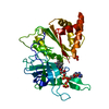
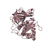
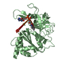
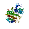
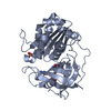
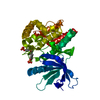
- PDB-1frn: THE INVOLVEMENT OF SER96 IN THE CATALYTIC MECHANISM OF FERREDOXIN... -
+ Open data

- Basic information
| Entry | Database: PDB / ID: 1frn | ||||||
|---|---|---|---|---|---|---|---|
| Title | THE INVOLVEMENT OF SER96 IN THE CATALYTIC MECHANISM OF FERREDOXIN-NADP+ REDUCTASE: STRUCTURE-FUNCTION RELATIONSHIP AS STUDIED BY SITE-DIRECTED MUTAGENESIS AND X-RAY CRYSTALLOGRAPHY | ||||||
 Components Components | FERREDOXIN-NADP+ REDUCTASE | ||||||
 Keywords Keywords | OXIDOREDUCTASE (NADP+(A) / FERREDOXIN(A)) | ||||||
| Function / homology |  Function and homology information Function and homology informationchloroplast thylakoid membrane protein complex / ferredoxin-NADP+ reductase / ferredoxin-NADP+ reductase activity / chloroplast stroma / photosynthesis / electron transport chain / electron transfer activity Similarity search - Function | ||||||
| Biological species |  Spinacia oleracea (spinach) Spinacia oleracea (spinach) | ||||||
| Method |  X-RAY DIFFRACTION / Resolution: 2 Å X-RAY DIFFRACTION / Resolution: 2 Å | ||||||
 Authors Authors | Bruns, C.M. / Karplus, P.A. | ||||||
 Citation Citation | Journal: Biochemistry / Year: 1995 Title: Involvement of serine 96 in the catalytic mechanism of ferredoxin-NADP+ reductase: structure--function relationship as studied by site-directed mutagenesis and X-ray crystallography. Authors: Aliverti, A. / Bruns, C.M. / Pandini, V.E. / Karplus, P.A. / Vanoni, M.A. / Curti, B. / Zanetti, G. #1: Journal: J.Mol.Biol. / Year: 1995Title: Refined Crystal Structure of Spinach Ferredoxin Reductase at 1.7 Angstroms Resolution: Oxidized, Reduced, and 2'-Phospho-5'-AMP Bound States Authors: Bruns, C.M. / Karplus, P.A. #2: Journal: Science / Year: 1991Title: Atomic Structure of Ferredoxin-Nadp+ Reductase: Prototype for a Structurally Novel Flavoenzyme Family Authors: Karplus, P.A. / Daniels, M.J. / Herriott, J.R. | ||||||
| History |
|
- Structure visualization
| Structure viewer | Molecule: MolmilJmol/JSmol |
|---|
- Downloads & links
-Download
| PDBx/mmCIF format | 1frn.cif.gz | 79.4 KB | Display | PDBx/mmCIF format |
|---|---|---|---|---|
| PDB format | pdb1frn.ent.gz | 58.3 KB | Display | PDB format |
| PDBx/mmJSON format | 1frn.json.gz | Tree view | PDBx/mmJSON format | |
| Others |  Other downloads Other downloads |
-Validation report
| Arichive directory | https://data.pdbj.org/pub/pdb/validation_reports/fr/1frnftp://data.pdbj.org/pub/pdb/validation_reports/fr/1frn | HTTPS FTP |
|---|
-Related structure data
| Similar structure data |
|---|


-Links
 PDBj
PDBj

- Assembly
| Deposited unit | 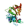
| ||||||||
|---|---|---|---|---|---|---|---|---|---|
| 1 |
| ||||||||
| Unit cell |
| ||||||||
| Atom site foot note | 1: CIS PROLINE - PRO 150 | ||||||||
| Components on special symmetry positions |
|
-Components
| #1: Protein | Mass: 35441.797 Da / Num. of mol.: 1 Source method: isolated from a genetically manipulated source Source: (gene. exp.) Spinacia oleracea (spinach) / Organ: LEAF / Plasmid: PDS12/RBSII SPH(I) / Production host:  |
|---|---|
| #2: Chemical | ChemComp-PO4 /   Mass: 94.971 Da / Num. of mol.: 1 / Source method: obtained synthetically / Formula: PO4 Mass: 94.971 Da / Num. of mol.: 1 / Source method: obtained synthetically / Formula: PO4 |
| #3: Chemical | ChemComp-SO4 /   Mass: 96.063 Da / Num. of mol.: 1 / Source method: obtained synthetically / Formula: SO4 Mass: 96.063 Da / Num. of mol.: 1 / Source method: obtained synthetically / Formula: SO4 |
| #4: Chemical | ChemComp-FAD /   Mass: 785.550 Da / Num. of mol.: 1 / Source method: obtained synthetically / Formula: C27H33N9O15P2 / Comment: FAD*YM Mass: 785.550 Da / Num. of mol.: 1 / Source method: obtained synthetically / Formula: C27H33N9O15P2 / Comment: FAD*YM |
| #5: Water | ChemComp-HOH /  Mass: 18.015 Da / Num. of mol.: 221 / Source method: isolated from a natural source / Formula: H2O Mass: 18.015 Da / Num. of mol.: 221 / Source method: isolated from a natural source / Formula: H2O |
| Compound details | COMPND PH 4.6, RESIDUE 96 MUTATED FROM SERINE TO VALINE, RECOMBINANT VARIANT WITH PHENYLALANINE AT ...COMPND PH 4.6, RESIDUE 96 MUTATED FROM SERINE TO VALINE, RECOMBINAN |
-Experimental details
-Experiment
| Experiment | Method: X-RAY DIFFRACTION |
|---|
- Sample preparation
| Crystal | Density Matthews: 2.47 Å3/Da / Density % sol: 50.29 % | ||||||||||||||||||||
|---|---|---|---|---|---|---|---|---|---|---|---|---|---|---|---|---|---|---|---|---|---|
| Crystal grow | *PLUS Temperature: 4 ℃ / pH: 4.5 / Method: unknown | ||||||||||||||||||||
| Components of the solutions | *PLUS
|
-Data collection
| Diffraction source | Wavelength: 1.5 |
|---|---|
| Detector | Type: SDMS / Date: Jun 15, 1994 |
| Radiation | Scattering type: x-ray |
| Radiation wavelength | Wavelength: 1.5 Å / Relative weight: 1 |
| Reflection | Redundancy: 4.8 % / Rmerge(I) obs: 0.088 |
| Reflection | *PLUS Num. obs: 22757 / Observed criterion σ(I): 0 / Rmerge(I) obs: 0.088 |
- Processing
| Software |
| ||||||||||||||||||||||||||||||
|---|---|---|---|---|---|---|---|---|---|---|---|---|---|---|---|---|---|---|---|---|---|---|---|---|---|---|---|---|---|---|---|
| Refinement | Highest resolution: 2 Å / σ(F): 0 /
| ||||||||||||||||||||||||||||||
| Refinement step | Cycle: LAST / Highest resolution: 2 Å
| ||||||||||||||||||||||||||||||
| Refine LS restraints |
|
