 Movie
Movie Controller


+ Open data

- Basic information
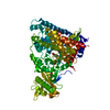
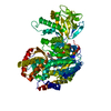
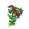
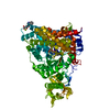
| Entry | Database: PDB / ID: 1fok | ||||||
|---|---|---|---|---|---|---|---|
| Title | STRUCTURE OF RESTRICTION ENDONUCLEASE FOKI BOUND TO DNA | ||||||
 Components Components |
| ||||||
 Keywords Keywords | HYDROLASE/DNA / COMPLEX (ENDONUCLEASE-DNA) / TYPE IIS / RESTRICTION ENDONUCLEASE / DEOXYRIBONUCLEASE / DNA HYDROLYSIS / DNA CLEAVAGE / HYDROLASE-DNA COMPLEX | ||||||
| Function / homology |  Function and homology information Function and homology informationtype II site-specific deoxyribonuclease / type II site-specific deoxyribonuclease activity / DNA restriction-modification system / DNA binding Similarity search - Function | ||||||
| Biological species |  Planomicrobium okeanokoites (bacteria) Planomicrobium okeanokoites (bacteria) | ||||||
| Method |  X-RAY DIFFRACTION / SYNCHROTRON / MIRAS / Resolution: 2.8 Å X-RAY DIFFRACTION / SYNCHROTRON / MIRAS / Resolution: 2.8 Å | ||||||
 Authors Authors | Aggarwal, D.A. / Wah, J.A. / Hirsch, L.F. / Dorner, I. / Schildkraut, A.K. | ||||||
 Citation Citation | Journal: Nature / Year: 1997 Title: Structure of the multimodular endonuclease FokI bound to DNA. Authors: Wah, D.A. / Hirsch, J.A. / Dorner, L.F. / Schildkraut, I. / Aggarwal, A.K. #1: Journal: FEBS Lett. / Year: 1997Title: Crystallization and Preliminary X-Ray Analysis of Restriction Endonuclease Foki Bound to DNA Authors: Hirsch, J.A. / Wah, D.A. / Dorner, L.F. / Schildkraut, I. / Aggarwal, A.K. | ||||||
| History |
|
- Structure visualization
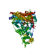
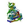
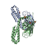
| Structure viewer | Molecule: MolmilJmol/JSmol |
|---|
- Downloads & links
-Download
| PDBx/mmCIF format | 1fok.cif.gz | 151.8 KB | Display | PDBx/mmCIF format |
|---|---|---|---|---|
| PDB format | pdb1fok.ent.gz | 115.2 KB | Display | PDB format |
| PDBx/mmJSON format | 1fok.json.gz | Tree view | PDBx/mmJSON format | |
| Others |  Other downloads Other downloads |
-Validation report
| Arichive directory | https://data.pdbj.org/pub/pdb/validation_reports/fo/1fokftp://data.pdbj.org/pub/pdb/validation_reports/fo/1fok | HTTPS FTP |
|---|
-Related structure data
| Similar structure data |
|---|


-Links
 PDBj
PDBj


- Assembly
| Deposited unit | 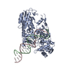
| ||||||||
|---|---|---|---|---|---|---|---|---|---|
| 1 |
| ||||||||
| Unit cell |
|
-Components
| #1: DNA chain | Mass: 6158.004 Da / Num. of mol.: 1 Source method: isolated from a genetically manipulated source Source: (gene. exp.) Planomicrobium okeanokoites (bacteria) / Strain: IFO12536 / Gene: FOKI / Production host: |
|---|---|
| #2: DNA chain | Mass: 6108.965 Da / Num. of mol.: 1 Source method: isolated from a genetically manipulated source |
| #3: Protein | Mass: 65519.230 Da / Num. of mol.: 1 Source method: isolated from a genetically manipulated source Source: (gene. exp.) Planomicrobium okeanokoites (bacteria) / References: UniProt: P14870 |
| #4: Water | ChemComp-HOH /  Mass: 18.015 Da / Num. of mol.: 172 / Source method: isolated from a natural source / Formula: H2O Mass: 18.015 Da / Num. of mol.: 172 / Source method: isolated from a natural source / Formula: H2O |
-Experimental details
-Experiment
| Experiment | Method: X-RAY DIFFRACTION / Number of used crystals: 7 |
|---|
- Sample preparation
| Crystal | Density Matthews: 3.51 Å3/Da / Density % sol: 62 % / Description: CRYSTALS WERE FLASH FROZEN IN NITROGEN STREAM. | ||||||||||||||||||||||||||||||||||||||||||||||||||||||||||||||||||||||||||||||
|---|---|---|---|---|---|---|---|---|---|---|---|---|---|---|---|---|---|---|---|---|---|---|---|---|---|---|---|---|---|---|---|---|---|---|---|---|---|---|---|---|---|---|---|---|---|---|---|---|---|---|---|---|---|---|---|---|---|---|---|---|---|---|---|---|---|---|---|---|---|---|---|---|---|---|---|---|---|---|---|
| Crystal grow | pH: 6 / Details: pH 6.00 | ||||||||||||||||||||||||||||||||||||||||||||||||||||||||||||||||||||||||||||||
| Crystal | *PLUS Density % sol: 62 % | ||||||||||||||||||||||||||||||||||||||||||||||||||||||||||||||||||||||||||||||
| Crystal grow | *PLUS pH: 6 / Method: vapor diffusion, hanging drop / Details: macroseeding | ||||||||||||||||||||||||||||||||||||||||||||||||||||||||||||||||||||||||||||||
| Components of the solutions | *PLUS
|
-Data collection
| Diffraction | Mean temperature: 130 K |
|---|---|
| Diffraction source | Source: SYNCHROTRON / Site: CHESS  / Beamline: F1 / Beamline: F1 |
| Detector | Type: SOL GRUNER, FUJI / Detector: CCD / Date: May 3, 1994 / Details: MIRRORS |
| Radiation | Monochromator: BENT, TRIANGULAR SI(111) / Monochromatic (M) / Laue (L): M / Scattering type: x-ray |
| Radiation wavelength | Relative weight: 1 |
| Reflection | Resolution: 2.8→100 Å / Num. obs: 26253 / % possible obs: 98.5 % / Observed criterion σ(I): 2 / Redundancy: 7.7 % / Biso Wilson estimate: 57.6 Å2 / Rmerge(I) obs: 0.075 |
| Reflection shell | Resolution: 2.8→2.9 Å / Redundancy: 2.7 % / Rmerge(I) obs: 0.325 / % possible all: 96.1 |
| Reflection | *PLUS Highest resolution: 2.8 Å / Lowest resolution: 100 Å / % possible obs: 98.5 % |
| Reflection shell | *PLUS Highest resolution: 2.8 Å / Lowest resolution: 2.9 Å / % possible obs: 96.1 % |
- Processing
| Software |
| ||||||||||||||||||||||||||||||||||||||||||||||||||||||||||||||||||||||||||||||||
|---|---|---|---|---|---|---|---|---|---|---|---|---|---|---|---|---|---|---|---|---|---|---|---|---|---|---|---|---|---|---|---|---|---|---|---|---|---|---|---|---|---|---|---|---|---|---|---|---|---|---|---|---|---|---|---|---|---|---|---|---|---|---|---|---|---|---|---|---|---|---|---|---|---|---|---|---|---|---|---|---|---|
| Refinement | Method to determine structure: MIRAS / Resolution: 2.8→8 Å / Rfactor Rfree error: 0.011 / Data cutoff high absF: 100000 / Data cutoff low absF: 0.1 / Isotropic thermal model: RESTRAINED / Cross valid method: THROUGHOUT / σ(F): 2
| ||||||||||||||||||||||||||||||||||||||||||||||||||||||||||||||||||||||||||||||||
| Displacement parameters | Biso mean: 38.2 Å2
| ||||||||||||||||||||||||||||||||||||||||||||||||||||||||||||||||||||||||||||||||
| Refine analyze |
| ||||||||||||||||||||||||||||||||||||||||||||||||||||||||||||||||||||||||||||||||
| Refinement step | Cycle: LAST / Resolution: 2.8→8 Å
| ||||||||||||||||||||||||||||||||||||||||||||||||||||||||||||||||||||||||||||||||
| Refine LS restraints |
| ||||||||||||||||||||||||||||||||||||||||||||||||||||||||||||||||||||||||||||||||
| LS refinement shell | Resolution: 2.8→2.92 Å / Rfactor Rfree error: 0.04 / Total num. of bins used: 8
| ||||||||||||||||||||||||||||||||||||||||||||||||||||||||||||||||||||||||||||||||
| Xplor file |
| ||||||||||||||||||||||||||||||||||||||||||||||||||||||||||||||||||||||||||||||||
| Software | *PLUS Name: X-PLOR / Version: 3.851 / Classification: refinement | ||||||||||||||||||||||||||||||||||||||||||||||||||||||||||||||||||||||||||||||||
| Refinement | *PLUS Highest resolution: 2.8 Å / Lowest resolution: 8 Å / σ(F): 2 | ||||||||||||||||||||||||||||||||||||||||||||||||||||||||||||||||||||||||||||||||
| Solvent computation | *PLUS | ||||||||||||||||||||||||||||||||||||||||||||||||||||||||||||||||||||||||||||||||
| Displacement parameters | *PLUS | ||||||||||||||||||||||||||||||||||||||||||||||||||||||||||||||||||||||||||||||||
| Refine LS restraints | *PLUS
| ||||||||||||||||||||||||||||||||||||||||||||||||||||||||||||||||||||||||||||||||
| LS refinement shell | *PLUS Rfactor obs: 0.354 |
