 Movie
Movie Controller


+ Open data

- Basic information
| Entry | Database: PDB / ID: 1fhe | ||||||
|---|---|---|---|---|---|---|---|
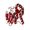
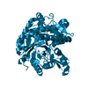
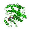
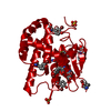
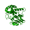
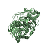
| Title | GLUTATHIONE TRANSFERASE (FH47) FROM FASCIOLA HEPATICA | ||||||
 Components Components | GLUTATHIONE TRANSFERASE | ||||||
 Keywords Keywords | TRANSFERASE / DETOXIFICATION / GLUTATHIONE TRANSFERASE | ||||||
| Function / homology |  Function and homology information Function and homology informationglutathione transferase / glutathione transferase activity / glutathione metabolic process / cytoplasm Similarity search - Function | ||||||
| Biological species |  Fasciola hepatica (liver fluke) Fasciola hepatica (liver fluke) | ||||||
| Method |  X-RAY DIFFRACTION / MOLECULAR REPLACEMENT / Resolution: 3 Å X-RAY DIFFRACTION / MOLECULAR REPLACEMENT / Resolution: 3 Å | ||||||
 Authors Authors | Rossjohn, J. / Parker, M.W. | ||||||
 Citation Citation | Journal: J.Mol.Biol. / Year: 1997 Title: Crystallization, structural determination and analysis of a novel parasite vaccine candidate: Fasciola hepatica glutathione S-transferase. Authors: Rossjohn, J. / Feil, S.C. / Wilce, M.C. / Sexton, J.L. / Spithill, T.W. / Parker, M.W. | ||||||
| History |
|
- Structure visualization
| Structure viewer | Molecule: MolmilJmol/JSmol |
|---|
- Downloads & links
-Download
| PDBx/mmCIF format | 1fhe.cif.gz | 53.1 KB | Display | PDBx/mmCIF format |
|---|---|---|---|---|
| PDB format | pdb1fhe.ent.gz | 39 KB | Display | PDB format |
| PDBx/mmJSON format | 1fhe.json.gz | Tree view | PDBx/mmJSON format | |
| Others |  Other downloads Other downloads |
-Validation report
| Arichive directory | https://data.pdbj.org/pub/pdb/validation_reports/fh/1fheftp://data.pdbj.org/pub/pdb/validation_reports/fh/1fhe | HTTPS FTP |
|---|
-Related structure data


-Links
 PDBj
PDBj

- Assembly
| Deposited unit | 
| ||||||||
|---|---|---|---|---|---|---|---|---|---|
| 1 | 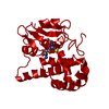
| ||||||||
| Unit cell |
|
-Components
| #1: Protein | Mass: 25318.242 Da / Num. of mol.: 1 Source method: isolated from a genetically manipulated source Source: (gene. exp.) Fasciola hepatica (liver fluke)Description: SECRETED AS A MONOMER, FORMS MEMBRANE-BOUND OLIGOMERS Cellular location: CYTOPLASM / Organ: LIVER / Production host:  |
|---|---|
| #2: Chemical | ChemComp-GSH /   Mass: 307.323 Da / Num. of mol.: 1 / Source method: obtained synthetically / Formula: C10H17N3O6S Mass: 307.323 Da / Num. of mol.: 1 / Source method: obtained synthetically / Formula: C10H17N3O6S |
| #3: Water | ChemComp-HOH /  Mass: 18.015 Da / Num. of mol.: 30 / Source method: isolated from a natural source / Formula: H2O Mass: 18.015 Da / Num. of mol.: 30 / Source method: isolated from a natural source / Formula: H2O |
-Experimental details
-Experiment
| Experiment | Method: X-RAY DIFFRACTION / Number of used crystals: 1 |
|---|
- Sample preparation
| Crystal | Density Matthews: 4.6 Å3/Da / Density % sol: 75 % | |||||||||||||||||||||||||
|---|---|---|---|---|---|---|---|---|---|---|---|---|---|---|---|---|---|---|---|---|---|---|---|---|---|---|
| Crystal grow | pH: 8.4 / Details: AMMONIUM SULFATE, GLUTATHIONE, TRIS PH 8.4 | |||||||||||||||||||||||||
| Crystal | *PLUS Density % sol: 52 % | |||||||||||||||||||||||||
| Crystal grow | *PLUS Temperature: 22 ℃ / Method: vapor diffusion, hanging drop | |||||||||||||||||||||||||
| Components of the solutions | *PLUS
|
-Data collection
| Diffraction | Mean temperature: 100 K |
|---|---|
| Diffraction source | Source: ROTATING ANODE / Type: RIGAKU / Wavelength: 1.5418 |
| Detector | Type: MARRESEARCH / Detector: IMAGE PLATE / Date: Oct 1, 1995 |
| Radiation | Monochromator: GRAPHITE(002) / Monochromatic (M) / Laue (L): M / Scattering type: x-ray |
| Radiation wavelength | Wavelength: 1.5418 Å / Relative weight: 1 |
| Reflection | Resolution: 3→30 Å / Num. obs: 9266 / % possible obs: 94.3 % / Rsym value: 0.11 |
| Reflection shell | Resolution: 3→3.1 Å / Rsym value: 0.39 |
| Reflection | *PLUS Rmerge(I) obs: 0.112 |
| Reflection shell | *PLUS Rmerge(I) obs: 0.396 |
- Processing
| Software |
| ||||||||||||||||||||||||||||||||||||||||||||||||||||||||||||
|---|---|---|---|---|---|---|---|---|---|---|---|---|---|---|---|---|---|---|---|---|---|---|---|---|---|---|---|---|---|---|---|---|---|---|---|---|---|---|---|---|---|---|---|---|---|---|---|---|---|---|---|---|---|---|---|---|---|---|---|---|---|
| Refinement | Method to determine structure: MOLECULAR REPLACEMENT Starting model: SCHISTOSOMA JAPONICUM GST Resolution: 3→30 Å / Data cutoff high absF: 10000000 / Data cutoff low absF: 0.001 / Cross valid method: THROUGHOUT / σ(F): 0
| ||||||||||||||||||||||||||||||||||||||||||||||||||||||||||||
| Refinement step | Cycle: LAST / Resolution: 3→30 Å
| ||||||||||||||||||||||||||||||||||||||||||||||||||||||||||||
| Refine LS restraints |
| ||||||||||||||||||||||||||||||||||||||||||||||||||||||||||||
| LS refinement shell | Resolution: 3→3.1 Å / Rfactor Rfree: 0.374 / Rfactor Rwork: 0.294 / Total num. of bins used: 8 | ||||||||||||||||||||||||||||||||||||||||||||||||||||||||||||
| Software | *PLUS Name: X-PLOR / Version: 3.1 / Classification: refinement | ||||||||||||||||||||||||||||||||||||||||||||||||||||||||||||
| Refine LS restraints | *PLUS
|
