 Movie
Movie Controller


+ Open data

- Basic information
| Entry | Database: PDB / ID: 1fc5 | |||||||||
|---|---|---|---|---|---|---|---|---|---|---|
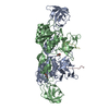
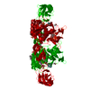
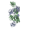
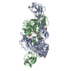
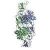
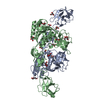
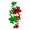
| Title | CRYSTAL STRUCTURE OF MOLYBDOPTERIN BIOSYNTHESIS MOEA PROTEIN | |||||||||
 Components Components | MOLYBDOPTERIN BIOSYNTHESIS MOEA PROTEIN | |||||||||
 Keywords Keywords | BIOSYNTHETIC PROTEIN / molybdopterin / four modules / with magnesium / Montreal-Kingston Bacterial Structural Genomics Initiative / BSGI / Structural Genomics | |||||||||
| Function / homology |  Function and homology information Function and homology informationmolybdopterin molybdotransferase activity / molybdopterin molybdotransferase / Mo-molybdopterin cofactor biosynthetic process / protein homodimerization activity / metal ion binding / identical protein binding / cytoplasm / cytosol Similarity search - Function | |||||||||
| Biological species |  | |||||||||
| Method |  X-RAY DIFFRACTION / SYNCHROTRON / Resolution: 2.2 Å X-RAY DIFFRACTION / SYNCHROTRON / Resolution: 2.2 Å | |||||||||
 Authors Authors | Huang, W. / Cygler, M. / Montreal-Kingston Bacterial Structural Genomics Initiative (BSGI) | |||||||||
 Citation Citation | Journal: J.Mol.Biol. / Year: 2001 Title: The crystal structure of Escherichia coli MoeA, a protein from the molybdopterin synthesis pathway. Authors: Schrag, J.D. / Huang, W. / Sivaraman, J. / Smith, C. / Plamondon, J. / Larocque, R. / Matte, A. / Cygler, M. | |||||||||
| History |
|
- Structure visualization
| Structure viewer | Molecule: MolmilJmol/JSmol |
|---|
- Downloads & links
-Download
| PDBx/mmCIF format | 1fc5.cif.gz | 171.1 KB | Display | PDBx/mmCIF format |
|---|---|---|---|---|
| PDB format | pdb1fc5.ent.gz | 135.6 KB | Display | PDB format |
| PDBx/mmJSON format | 1fc5.json.gz | Tree view | PDBx/mmJSON format | |
| Others |  Other downloads Other downloads |
-Validation report
| Arichive directory | https://data.pdbj.org/pub/pdb/validation_reports/fc/1fc5ftp://data.pdbj.org/pub/pdb/validation_reports/fc/1fc5 | HTTPS FTP |
|---|
-Related structure data
| Similar structure data | |
|---|---|
| Other databases |


-Links
 PDBj
PDBj- Assembly
| Deposited unit | 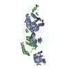
| ||||||||
|---|---|---|---|---|---|---|---|---|---|
| 1 |
| ||||||||
| Unit cell |
| ||||||||
| Details | The biological assembly is a dimer constructed from chain A a symmetry partner generated by the two-fold. |
-Components
| #1: Protein | Mass: 44481.215 Da / Num. of mol.: 2 Source method: isolated from a genetically manipulated source Source: (gene. exp.) #2: Chemical |   Mass: 24.305 Da / Num. of mol.: 2 / Source method: obtained synthetically / Formula: Mg Mass: 24.305 Da / Num. of mol.: 2 / Source method: obtained synthetically / Formula: Mg#3: Water | ChemComp-HOH / |  Mass: 18.015 Da / Num. of mol.: 500 / Source method: isolated from a natural source / Formula: H2O Mass: 18.015 Da / Num. of mol.: 500 / Source method: isolated from a natural source / Formula: H2OHas protein modification | Y | |
|---|
-Experimental details
-Experiment
| Experiment | Method: X-RAY DIFFRACTION / Number of used crystals: 1 |
|---|
- Sample preparation
| Crystal | Density Matthews: 2.27 Å3/Da / Density % sol: 45.76 % | ||||||||||||||||||||||||||||||||||||||||||||||||||||||
|---|---|---|---|---|---|---|---|---|---|---|---|---|---|---|---|---|---|---|---|---|---|---|---|---|---|---|---|---|---|---|---|---|---|---|---|---|---|---|---|---|---|---|---|---|---|---|---|---|---|---|---|---|---|---|---|
| Crystal grow | Temperature: 293 K / Method: vapor diffusion / pH: 6.5 Details: PEG8000, Cacodylate, pH 6.5, VAPOR DIFFUSION, temperature 20K | ||||||||||||||||||||||||||||||||||||||||||||||||||||||
| Crystal | *PLUS Density % sol: 44 % | ||||||||||||||||||||||||||||||||||||||||||||||||||||||
| Crystal grow | *PLUS pH: 7.5 / Method: vapor diffusion, hanging drop / Details: used macroseeding | ||||||||||||||||||||||||||||||||||||||||||||||||||||||
| Components of the solutions | *PLUS
|
-Data collection
| Diffraction | Mean temperature: 100 K |
|---|---|
| Diffraction source | Source: SYNCHROTRON / Site: NSLS  / Beamline: X8C / Wavelength: 0.9795 / Beamline: X8C / Wavelength: 0.9795 |
| Detector | Type: ADSC QUANTUM 4 / Detector: CCD / Date: May 28, 2000 |
| Radiation | Protocol: SINGLE WAVELENGTH / Monochromatic (M) / Laue (L): M / Scattering type: x-ray |
| Radiation wavelength | Wavelength: 0.9795 Å / Relative weight: 1 |
| Reflection | Resolution: 2.2→50 Å / Num. all: 41012 / Num. obs: 231130 / % possible obs: 98.2 % / Observed criterion σ(F): 0 / Observed criterion σ(I): 0 / Redundancy: 5.8 % / Biso Wilson estimate: 40 Å2 / Rmerge(I) obs: 0.059 / Net I/σ(I): 19.6 |
| Reflection shell | Resolution: 2.2→2.23 Å / Redundancy: 2.1 % / Rmerge(I) obs: 0.255 / % possible all: 88.3 |
| Reflection | *PLUS Lowest resolution: 20 Å / Num. obs: 40862 / Num. measured all: 232716 / Rmerge(I) obs: 0.067 |
| Reflection shell | *PLUS % possible obs: 88.3 % / Rmerge(I) obs: 0.209 / Mean I/σ(I) obs: 12.6 |
- Processing
| Software |
| ||||||||||||||||||||
|---|---|---|---|---|---|---|---|---|---|---|---|---|---|---|---|---|---|---|---|---|---|
| Refinement | Resolution: 2.2→50 Å / σ(F): 0 / σ(I): 0 / Stereochemistry target values: Engh & Huber
| ||||||||||||||||||||
| Refinement step | Cycle: LAST / Resolution: 2.2→50 Å
| ||||||||||||||||||||
| Refine LS restraints |
| ||||||||||||||||||||
| Software | *PLUS Name: CNS / Classification: refinement | ||||||||||||||||||||
| Refinement | *PLUS Lowest resolution: 50 Å / σ(F): 0 / Rfactor obs: 0.226 / Rfactor Rfree: 0.277 | ||||||||||||||||||||
| Solvent computation | *PLUS | ||||||||||||||||||||
| Displacement parameters | *PLUS |
