 Movie
Movie Controller

[English] 日本語
 Yorodumi
- PDB-1f38: X-RAY CRYSTALLOGRAPHIC STRUCTURE OF PRECORRIN 8W DECARBOXYLASE, T... -
+ Open data

- Basic information
| Entry | Database: PDB / ID: 1f38 | ||||||
|---|---|---|---|---|---|---|---|
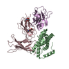
| Title | X-RAY CRYSTALLOGRAPHIC STRUCTURE OF PRECORRIN 8W DECARBOXYLASE, THE PRODUCT OF GENE MT0146 IN THE METHANOBACTERIUM THERMOAUTOTROPHICUM GENOME | ||||||
 Components Components | PRECORRIN-8W DECARBOXYLASE | ||||||
 Keywords Keywords | LYASE / Decarboxylase / Structural Genomics / PSI / Protein Structure Initiative / Northeast Structural Genomics Consortium / NESG | ||||||
| Function / homology |  Function and homology information Function and homology informationcobalt-precorrin-6B (C15)-methyltransferase [decarboxylating] / cobalt-precorrin-6B C5-methyltransferase activity / : / protein methyltransferase activity / methylation Similarity search - Function | ||||||
| Biological species |   Methanothermobacter thermautotrophicus (archaea) Methanothermobacter thermautotrophicus (archaea) | ||||||
| Method |  X-RAY DIFFRACTION / SYNCHROTRON / MAD / Resolution: 2.4 Å X-RAY DIFFRACTION / SYNCHROTRON / MAD / Resolution: 2.4 Å | ||||||
 Authors Authors | Keller, J.P. / Smith, P.M. / Hunt, J.F. / Northeast Structural Genomics Consortium (NESG) | ||||||
 Citation Citation | Journal: Structure / Year: 2002 Title: The crystal structure of MT0146/CbiT suggests that the putative precorrin-8w decarboxylase is a methyltransferase Authors: Keller, J.P. / Smith, P.M. / Benach, J. / Christendat, D. / deTitta, G.T. / Hunt, J.F. | ||||||
| History |
|
- Structure visualization
| Structure viewer | Molecule: MolmilJmol/JSmol |
|---|
- Downloads & links
-Download
| PDBx/mmCIF format | 1f38.cif.gz | 154.5 KB | Display | PDBx/mmCIF format |
|---|---|---|---|---|
| PDB format | pdb1f38.ent.gz | 122.5 KB | Display | PDB format |
| PDBx/mmJSON format | 1f38.json.gz | Tree view | PDBx/mmJSON format | |
| Others |  Other downloads Other downloads |
-Validation report
| Arichive directory | https://data.pdbj.org/pub/pdb/validation_reports/f3/1f38ftp://data.pdbj.org/pub/pdb/validation_reports/f3/1f38 | HTTPS FTP |
|---|
-Related structure data
| Related structure data |  1kxzC  1l3bC  1l3cC  1l3iC C: citing same article ( |
|---|---|
| Similar structure data | |
| Other databases |


-Links
 PDBj
PDBj- Assembly
| Deposited unit | 
| ||||||||
|---|---|---|---|---|---|---|---|---|---|
| 1 |
| ||||||||
| Unit cell |
| ||||||||
| Details | The biological assembly is a tetramer constructed from chains A -> D. |
-Components
| #1: Protein | Mass: 21112.131 Da / Num. of mol.: 4 Source method: isolated from a genetically manipulated source Source: (gene. exp.) Methanothermobacter thermautotrophicus (archaea)Description: GENOMIC DNA / Gene: MT0146 / Plasmid: PET14A DERIVATIVE / Production host:  #2: Water | ChemComp-HOH / |  Mass: 18.015 Da / Num. of mol.: 180 / Source method: isolated from a natural source / Formula: H2O Mass: 18.015 Da / Num. of mol.: 180 / Source method: isolated from a natural source / Formula: H2OHas protein modification | Y | |
|---|
-Experimental details
-Experiment
| Experiment | Method: X-RAY DIFFRACTION / Number of used crystals: 1 |
|---|
- Sample preparation
| Crystal | Density Matthews: 2.54 Å3/Da / Density % sol: 51.62 % |
|---|---|
| Crystal grow | Temperature: 299 K / Method: vapor diffusion, hanging drop Details: Magnesium Chloride, PEG 8000, VAPOR DIFFUSION, HANGING DROP, temperature 26K |
-Data collection
| Diffraction | Mean temperature: 130 K |
|---|---|
| Diffraction source | Source: SYNCHROTRON / Site: APS  / Beamline: 32-ID / Wavelength: 1.06884 / Beamline: 32-ID / Wavelength: 1.06884 |
| Detector | Type: MARRESEARCH / Detector: CCD / Date: May 19, 2000 |
| Radiation | Protocol: MAD / Monochromatic (M) / Laue (L): M / Scattering type: x-ray |
| Radiation wavelength | Wavelength: 1.06884 Å / Relative weight: 1 |
| Reflection | Resolution: 2.4→40 Å / Num. all: 54692 / Num. obs: 33548 / % possible obs: 73.8 % / Observed criterion σ(F): 1 / Observed criterion σ(I): 3 / Redundancy: 13.2 % / Biso Wilson estimate: 37.8 Å2 / Rmerge(I) obs: 0.127 / Net I/σ(I): 25 |
| Reflection shell | Resolution: 2.4→2.51 Å / Redundancy: 13.2 % / Rmerge(I) obs: 0.08 / % possible all: 91.7 |
- Processing
| Software |
| ||||||||||||||||||||
|---|---|---|---|---|---|---|---|---|---|---|---|---|---|---|---|---|---|---|---|---|---|
| Refinement | Method to determine structure: MAD / Resolution: 2.4→100 Å / σ(F): 1 / σ(I): 1 / Stereochemistry target values: X-PLOR 3.851
| ||||||||||||||||||||
| Refinement step | Cycle: LAST / Resolution: 2.4→100 Å
| ||||||||||||||||||||
| Refine LS restraints |
|
