 Movie
Movie Controller

[English] 日本語
 Yorodumi
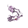
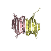
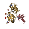
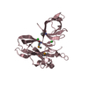
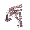
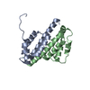
- PDB-1f2r: NMR STRUCTURE OF THE HETERODIMERIC COMPLEX BETWEEN CAD DOMAINS OF... -
+ Open data

- Basic information
| Entry | Database: PDB / ID: 1f2r | ||||||
|---|---|---|---|---|---|---|---|
| Title | NMR STRUCTURE OF THE HETERODIMERIC COMPLEX BETWEEN CAD DOMAINS OF CAD AND ICAD | ||||||
 Components Components |
| ||||||
 Keywords Keywords | DNA BINDING PROTEIN / ALPHA-BETA ROLL / PROTEIN-PROTEIN COMPLEX | ||||||
| Function / homology |  Function and homology information Function and homology informationApoptosis induced DNA fragmentation / deoxyribonuclease inhibitor activity / negative regulation of apoptotic DNA fragmentation / apoptotic chromosome condensation / DNA nuclease activity / Hydrolases / apoptotic DNA fragmentation / nuclease activity / negative regulation of execution phase of apoptosis / DNA catabolic process ...Apoptosis induced DNA fragmentation / deoxyribonuclease inhibitor activity / negative regulation of apoptotic DNA fragmentation / apoptotic chromosome condensation / DNA nuclease activity / Hydrolases / apoptotic DNA fragmentation / nuclease activity / negative regulation of execution phase of apoptosis / DNA catabolic process / thymocyte apoptotic process / protein folding chaperone / DNA endonuclease activity / disordered domain specific binding / protein folding / positive regulation of apoptotic process / protein domain specific binding / hydrolase activity / apoptotic process / chromatin / nucleolus / enzyme binding / protein-containing complex / DNA binding / nucleoplasm / identical protein binding / nucleus / plasma membrane / cytosol Similarity search - Function | ||||||
| Biological species |  | ||||||
| Method | SOLUTION NMR / simulated annealing | ||||||
 Authors Authors | Otomo, T. / Sakahira, H. / Uegaki, K. / Nagata, S. / Yamazaki, T. | ||||||
 Citation Citation | Journal: Nat.Struct.Biol. / Year: 2000 Title: Structure of the heterodimeric complex between CAD domains of CAD and ICAD. Authors: Otomo, T. / Sakahira, H. / Uegaki, K. / Nagata, S. / Yamazaki, T. #1: Journal: J.Mol.Biol. / Year: 2000Title: Structure of the CAD Domain of Caspase-Activated DNase and Interaction with the CAD Domain of its Inhibitor. Authors: Uegaki, K. / Otomo, T. / Sakahira, H. / Shimizu, M. / Yumoto, N. / Kyogoku, Y. / Nagata, S. / Yamazaki, T. #2: Journal: Nature / Year: 1998Title: A Caspase-Activated DNase that Degrades DNA During Apoptosis, and its Inhibitor ICAD. Authors: Enari, M. / Sakahira, H. / Yokoyama, H. / Okawa, K. / Iwamatsu, A. / Nagata, S. #3: Journal: Nature / Year: 1998Title: Cleavage of CAD Inhibitor in CAD Activation and DNA Degradation During Apoptosis. Authors: Sakahira, H. / Enari, M. / Nagata, S. | ||||||
| History |
|
- Structure visualization
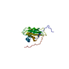
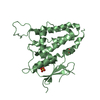
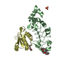
| Structure viewer | Molecule: MolmilJmol/JSmol |
|---|
- Downloads & links
-Download
| PDBx/mmCIF format | 1f2r.cif.gz | 74.7 KB | Display | PDBx/mmCIF format |
|---|---|---|---|---|
| PDB format | pdb1f2r.ent.gz | 57.4 KB | Display | PDB format |
| PDBx/mmJSON format | 1f2r.json.gz | Tree view | PDBx/mmJSON format | |
| Others |  Other downloads Other downloads |
-Validation report
| Arichive directory | https://data.pdbj.org/pub/pdb/validation_reports/f2/1f2rftp://data.pdbj.org/pub/pdb/validation_reports/f2/1f2r | HTTPS FTP |
|---|
-Related structure data
| Related structure data | |
|---|---|
| Similar structure data |

-Links
 PDBj
PDBj
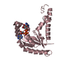
- Assembly
| Deposited unit | 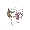
| |||||||||
|---|---|---|---|---|---|---|---|---|---|---|
| 1 |
| |||||||||
| NMR ensembles |
|
-Components
| #1: Protein | Mass: 9613.200 Da / Num. of mol.: 1 / Fragment: N-TERMINAL DOMAIN (CAD DOMAIN), RESIDUES 1-87 Source method: isolated from a genetically manipulated source Source: (gene. exp.)  |
|---|---|
| #2: Protein | Mass: 11076.416 Da / Num. of mol.: 1 / Fragment: N-TERMINAL DOMAIN (CAD DOMAIN), RESIDUES 1-100 Source method: isolated from a genetically manipulated source Source: (gene. exp.) |
-Experimental details
-Experiment
| Experiment | Method: SOLUTION NMR | ||||||||||||||||||||||||||||
|---|---|---|---|---|---|---|---|---|---|---|---|---|---|---|---|---|---|---|---|---|---|---|---|---|---|---|---|---|---|
| NMR experiment |
|
- Sample preparation
| Details |
| |||||||||||||||
|---|---|---|---|---|---|---|---|---|---|---|---|---|---|---|---|---|
| Sample conditions | Ionic strength: 0 / pH: 6.4 / Pressure: ambient / Temperature: 308 K | |||||||||||||||
| Crystal grow | *PLUS Method: other / Details: NMR |
-NMR measurement
| NMR spectrometer |
|
|---|
- Processing
| NMR software |
| ||||||||||||
|---|---|---|---|---|---|---|---|---|---|---|---|---|---|
| Refinement | Method: simulated annealing / Software ordinal: 1 Details: the structures are based on a total of 2646 restraints, 2503 are NOE-derived distance constraints, 93 dihedral angle restraints,50 distance restraints from hydrogen bonds. | ||||||||||||
| NMR ensemble | Conformers submitted total number: 1 |
 X-PLOR