 Movie
Movie Controller

[English] 日本語
 Yorodumi
- PDB-1ezz: CRYSTAL STRUCTURE OF E. COLI ASPARTATE TRANSCARBAMOYLASE P268A MU... -
+ Open data

- Basic information
| Entry | Database: PDB / ID: 1ezz | ||||||
|---|---|---|---|---|---|---|---|
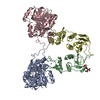
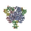
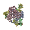
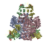
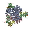
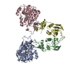
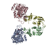
| Title | CRYSTAL STRUCTURE OF E. COLI ASPARTATE TRANSCARBAMOYLASE P268A MUTANT IN THE T-STATE | ||||||
 Components Components |
| ||||||
 Keywords Keywords | TRANSFERASE / aspartate transcarbamoylase / aspartate carbamoyltransferase / cis-proline / cis-amino acid | ||||||
| Function / homology |  Function and homology information Function and homology informationaspartate carbamoyltransferase complex / pyrimidine nucleotide biosynthetic process / aspartate carbamoyltransferase / aspartate carbamoyltransferase activity / L-glutamine metabolic process / amino acid binding / protein homotrimerization / 'de novo' UMP biosynthetic process / 'de novo' pyrimidine nucleobase biosynthetic process / zinc ion binding ...aspartate carbamoyltransferase complex / pyrimidine nucleotide biosynthetic process / aspartate carbamoyltransferase / aspartate carbamoyltransferase activity / L-glutamine metabolic process / amino acid binding / protein homotrimerization / 'de novo' UMP biosynthetic process / 'de novo' pyrimidine nucleobase biosynthetic process / zinc ion binding / identical protein binding / cytosol / cytoplasm Similarity search - Function | ||||||
| Biological species |  | ||||||
| Method |  X-RAY DIFFRACTION / Resolution: 2.7 Å X-RAY DIFFRACTION / Resolution: 2.7 Å | ||||||
 Authors Authors | Jin, L. / Stec, B. / Kantrowitz, E.R. | ||||||
 Citation Citation | Journal: Biochemistry / Year: 2000 Title: A cis-proline to alanine mutant of E. coli aspartate transcarbamoylase: kinetic studies and three-dimensional crystal structures. Authors: Jin, L. / Stec, B. / Kantrowitz, E.R. #1: Journal: Biochemistry / Year: 1990Title: Structural Consequences of Effector Binding to the T State of Aspartate Carbamoyltransferase: Crystal Structures of the Unligated and ATP- and CTP-complexed enzymes at 2.6-A resolution Authors: Stevens, R.C. / Gouaux, J.E. / Lipscomb, W.N. | ||||||
| History |
|
- Structure visualization
| Structure viewer | Molecule: MolmilJmol/JSmol |
|---|
- Downloads & links
-Download
| PDBx/mmCIF format | 1ezz.cif.gz | 194.4 KB | Display | PDBx/mmCIF format |
|---|---|---|---|---|
| PDB format | pdb1ezz.ent.gz | 154.9 KB | Display | PDB format |
| PDBx/mmJSON format | 1ezz.json.gz | Tree view | PDBx/mmJSON format | |
| Others |  Other downloads Other downloads |
-Validation report
| Arichive directory | https://data.pdbj.org/pub/pdb/validation_reports/ez/1ezzftp://data.pdbj.org/pub/pdb/validation_reports/ez/1ezz | HTTPS FTP |
|---|
-Related structure data


-Links
 PDBj
PDBj
- Assembly
| Deposited unit | 
| ||||||||
|---|---|---|---|---|---|---|---|---|---|
| 1 | 
| ||||||||
| Unit cell |
| ||||||||
| Components on special symmetry positions |
| ||||||||
| Details | The biological assembly is a dodecamer. The entire molecule requires two symmetry partners generated by rotations around the three-fold |
-Components
| #1: Protein | Mass: 34311.070 Da / Num. of mol.: 2 / Mutation: P268A Source method: isolated from a genetically manipulated source Source: (gene. exp.) Plasmid: PEK412, A PLASMID WITH THE PYRBI GENES INSERTED INTO PUC119 Production host: #2: Protein | Mass: 17143.625 Da / Num. of mol.: 2 Source method: isolated from a genetically manipulated source Source: (gene. exp.) Plasmid: PEK412, A PLASMID WITH THE PYRBI GENES INSERTED INTO PUC119 Production host: #3: Chemical |   Mass: 65.409 Da / Num. of mol.: 2 / Source method: obtained synthetically / Formula: Zn Mass: 65.409 Da / Num. of mol.: 2 / Source method: obtained synthetically / Formula: Zn#4: Water | ChemComp-HOH / |  Mass: 18.015 Da / Num. of mol.: 247 / Source method: isolated from a natural source / Formula: H2O Mass: 18.015 Da / Num. of mol.: 247 / Source method: isolated from a natural source / Formula: H2O |
|---|
-Experimental details
-Experiment
| Experiment | Method: X-RAY DIFFRACTION / Number of used crystals: 1 |
|---|
- Sample preparation
| Crystal | Density Matthews: 3.13 Å3/Da / Density % sol: 60.66 % | ||||||||||||||||||||
|---|---|---|---|---|---|---|---|---|---|---|---|---|---|---|---|---|---|---|---|---|---|
| Crystal grow | Temperature: 293 K / Method: vapor diffusion, hanging drop / pH: 7 Details: ENZYME: 24 mg/ml; 1:1 enzyme to-buffer ratio, BUFFER: 20 mm HEPES, 14% (w/v) PEG 1450, pH 7.0, VAPOR DIFFUSION, HANGING DROP, temperature 293.0K | ||||||||||||||||||||
| Crystal grow | *PLUS Temperature: 20 ℃ | ||||||||||||||||||||
| Components of the solutions | *PLUS
|
-Data collection
| Diffraction | Mean temperature: 295 K |
|---|---|
| Diffraction source | Source: ROTATING ANODE / Type: RIGAKU RU200 / Wavelength: 1.5418 |
| Detector | Type: UCSD MARK III / Detector: AREA DETECTOR / Date: Jun 19, 1998 |
| Radiation | Protocol: SINGLE WAVELENGTH / Monochromatic (M) / Laue (L): M / Scattering type: x-ray |
| Radiation wavelength | Wavelength: 1.5418 Å / Relative weight: 1 |
| Reflection | Resolution: 2.7→30 Å / Num. all: 34450 / Num. obs: 34422 / % possible obs: 99.9 % / Observed criterion σ(F): 0 / Observed criterion σ(I): 0 / Redundancy: 3.2 % / Biso Wilson estimate: 41.1 Å2 / Rmerge(I) obs: 0.073 / Net I/σ(I): 4.296 |
| Reflection shell | Resolution: 2.7→2.91 Å / Redundancy: 2.17 % / Rmerge(I) obs: 0.427 / Num. unique all: 13768 / % possible all: 99.8 |
- Processing
| Software |
| |||||||||||||||||||||||||
|---|---|---|---|---|---|---|---|---|---|---|---|---|---|---|---|---|---|---|---|---|---|---|---|---|---|---|
| Refinement | Resolution: 2.7→8 Å / σ(F): 2 / σ(I): 0 / Stereochemistry target values: Engh & Huber
| |||||||||||||||||||||||||
| Refinement step | Cycle: LAST / Resolution: 2.7→8 Å
| |||||||||||||||||||||||||
| Refine LS restraints |
| |||||||||||||||||||||||||
| Software | *PLUS Name: X-PLOR / Version: 3.1 / Classification: refinement | |||||||||||||||||||||||||
| Refinement | *PLUS Highest resolution: 2.7 Å / Lowest resolution: 8 Å / σ(F): 2 / Rfactor obs: 0.182 | |||||||||||||||||||||||||
| Solvent computation | *PLUS | |||||||||||||||||||||||||
| Displacement parameters | *PLUS | |||||||||||||||||||||||||
| Refine LS restraints | *PLUS
|
