 Movie
Movie Controller


+ Open data

- Basic information
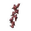
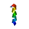
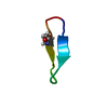
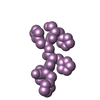
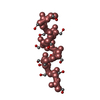
| Entry | Database: PDB / ID: 1ew1 | ||||||||||||||||||
|---|---|---|---|---|---|---|---|---|---|---|---|---|---|---|---|---|---|---|---|
| Title | RECA PROTEIN-BOUND SINGLE-STRANDED DNA | ||||||||||||||||||
 Components Components | DNA (5'-D(* Keywords KeywordsDNA / DEOXYRIBOSE-BASE STACKING / SINGLE-STRANDED DNA | Function / homology | DNA |  Function and homology information Function and homology informationMethod | SOLUTION NMR / simulated annealing |  Authors AuthorsNishinaka, T. / Ito, Y. / Yokoyama, S. / Shibata, T. |  CitationJournal: Proc.Natl.Acad.Sci.USA / Year: 1997 CitationJournal: Proc.Natl.Acad.Sci.USA / Year: 1997Title: An extended DNA structure through deoxyribose-base stacking induced by RecA protein. Authors: Nishinaka, T. / Ito, Y. / Yokoyama, S. / Shibata, T. History |
|
- Structure visualization
| Structure viewer | Molecule: MolmilJmol/JSmol |
|---|
- Downloads & links
-Download
| PDBx/mmCIF format | 1ew1.cif.gz | 27.9 KB | Display | PDBx/mmCIF format |
|---|---|---|---|---|
| PDB format | pdb1ew1.ent.gz | 18.1 KB | Display | PDB format |
| PDBx/mmJSON format | 1ew1.json.gz | Tree view | PDBx/mmJSON format | |
| Others |  Other downloads Other downloads |
-Validation report
| Arichive directory | https://data.pdbj.org/pub/pdb/validation_reports/ew/1ew1ftp://data.pdbj.org/pub/pdb/validation_reports/ew/1ew1 | HTTPS FTP |
|---|
-Related structure data


-Links
 PDBj
PDBj

- Assembly
| Deposited unit | 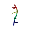
| |||||||||
|---|---|---|---|---|---|---|---|---|---|---|
| 1 |
| |||||||||
| NMR ensembles |
|
-Components
| #1: DNA chain | Mass: 1190.830 Da / Num. of mol.: 1 / Source method: obtained synthetically / Details: RECA PROTEIN-BOUND SINGLE-STRANDED DNA |
|---|
-Experimental details
-Experiment
| Experiment | Method: SOLUTION NMR |
|---|---|
| NMR experiment | Type: 2D NOESY |
| NMR details | Text: This structure was determined using transferred NOE techniques. THE ROOT-MEAN-SQUARE DEVIATION OF THE T-A-C REGION IS 0.30 ANGSTROM. THE FOURTH RESIDUE (G) IS DISORDERED BECAUSE OF FEW NOE CONSTRAINTS |
- Sample preparation
| Details | Contents: 0.8mM d(TpApCpG); 20mM Tris-Cl, 6.7mM MgCl2, 150mM NaCl Solvent system: D2O |
|---|---|
| Sample conditions | Ionic strength: 6.7mM MgCl2, 150mM NaCl / pH: 7.1 / Pressure: ambient / Temperature: 298 K |
| Crystal grow | *PLUS Method: other / Details: NMR |
-NMR measurement
| NMR spectrometer | Type: Bruker AMX / Manufacturer: Bruker / Model: AMX / Field strength: 600 MHz |
|---|
- Processing
| NMR software |
| ||||||||||||
|---|---|---|---|---|---|---|---|---|---|---|---|---|---|
| Refinement | Method: simulated annealing / Software ordinal: 1 | ||||||||||||
| NMR representative | Selection criteria: lowest energy | ||||||||||||
| NMR ensemble | Conformer selection criteria: structures with the lowest energy Conformers calculated total number: 100 / Conformers submitted total number: 10 |
 X-PLOR