 Movie
Movie Controller

[English] 日本語
 Yorodumi
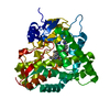
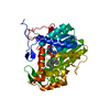
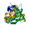
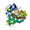
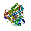
- PDB-1eup: X-RAY CRYSTAL STRUCTURE OF CYTOCHROME P450ERYF WITH ANDROSTENDION... -
+ Open data

- Basic information
| Entry | Database: PDB / ID: 1eup | |||||||||
|---|---|---|---|---|---|---|---|---|---|---|
| Title | X-RAY CRYSTAL STRUCTURE OF CYTOCHROME P450ERYF WITH ANDROSTENDIONE BOUND | |||||||||
 Components Components | CYTOCHROME P450ERYF | |||||||||
 Keywords Keywords | OXIDOREDUCTASE / cytochrome P450 / steroid / androstenedione / cytochrome P450eryF | |||||||||
| Function / homology |  Function and homology information Function and homology information6-deoxyerythronolide B hydroxylase / erythromycin biosynthetic process / oxidoreductase activity, acting on paired donors, with incorporation or reduction of molecular oxygen / monooxygenase activity / iron ion binding / heme binding / cytoplasm Similarity search - Function | |||||||||
| Biological species |  Saccharopolyspora erythraea (bacteria) Saccharopolyspora erythraea (bacteria) | |||||||||
| Method |  X-RAY DIFFRACTION / Resolution: 2.1 Å X-RAY DIFFRACTION / Resolution: 2.1 Å | |||||||||
 Authors Authors | Cupp-Vickery, J.R. / Anderson, R. / Hatziris, Z. | |||||||||
 Citation Citation | Journal: Proc.Natl.Acad.Sci.USA / Year: 2000 Title: Crystal structures of ligand complexes of P450eryF exhibiting homotropic cooperativity. Authors: Cupp-Vickery, J. / Anderson, R. / Hatziris, Z. | |||||||||
| History |
|
- Structure visualization
| Structure viewer | Molecule: MolmilJmol/JSmol |
|---|
- Downloads & links
-Download
| PDBx/mmCIF format | 1eup.cif.gz | 100.7 KB | Display | PDBx/mmCIF format |
|---|---|---|---|---|
| PDB format | pdb1eup.ent.gz | 75.2 KB | Display | PDB format |
| PDBx/mmJSON format | 1eup.json.gz | Tree view | PDBx/mmJSON format | |
| Others |  Other downloads Other downloads |
-Validation report
| Arichive directory | https://data.pdbj.org/pub/pdb/validation_reports/eu/1eupftp://data.pdbj.org/pub/pdb/validation_reports/eu/1eup | HTTPS FTP |
|---|
-Related structure data


-Links
 PDBj
PDBj

- Assembly
| Deposited unit | 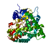
| ||||||||
|---|---|---|---|---|---|---|---|---|---|
| 1 |
| ||||||||
| Unit cell |
|
-Components
| #1: Protein | Mass: 44515.152 Da / Num. of mol.: 1 / Source method: isolated from a natural source / Source: (natural) Saccharopolyspora erythraea (bacteria) / References: UniProt: Q00441 | ||
|---|---|---|---|
| #2: Chemical | ChemComp-HEM /   Mass: 616.487 Da / Num. of mol.: 1 / Source method: obtained synthetically / Formula: C34H32FeN4O4 Mass: 616.487 Da / Num. of mol.: 1 / Source method: obtained synthetically / Formula: C34H32FeN4O4 | ||
| #3: Chemical | 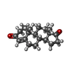  Mass: 286.409 Da / Num. of mol.: 2 / Source method: obtained synthetically / Formula: C19H26O2 Mass: 286.409 Da / Num. of mol.: 2 / Source method: obtained synthetically / Formula: C19H26O2#4: Water | ChemComp-HOH / |  Mass: 18.015 Da / Num. of mol.: 331 / Source method: isolated from a natural source / Formula: H2O Mass: 18.015 Da / Num. of mol.: 331 / Source method: isolated from a natural source / Formula: H2O |
-Experimental details
-Experiment
| Experiment | Method: X-RAY DIFFRACTION / Number of used crystals: 1 |
|---|
- Sample preparation
| Crystal | Density Matthews: 2.39 Å3/Da / Density % sol: 48.56 % | ||||||||||||||||||||||||||||||||||||||||||
|---|---|---|---|---|---|---|---|---|---|---|---|---|---|---|---|---|---|---|---|---|---|---|---|---|---|---|---|---|---|---|---|---|---|---|---|---|---|---|---|---|---|---|---|
| Crystal grow | Temperature: 295 K / Method: vapor diffusion, hanging drop / pH: 8.5 Details: PEG 4000 NaAc Tris, pH 8.5, VAPOR DIFFUSION, HANGING DROP, temperature 22K | ||||||||||||||||||||||||||||||||||||||||||
| Crystal grow | *PLUS Details: used to seeding | ||||||||||||||||||||||||||||||||||||||||||
| Components of the solutions | *PLUS
|
-Data collection
| Diffraction | Mean temperature: 22 K |
|---|---|
| Diffraction source | Source: ROTATING ANODE / Type: RIGAKU / Wavelength: 1.54 |
| Detector | Type: RIGAKU RAXIS II / Detector: IMAGE PLATE / Date: Jan 1, 1999 |
| Radiation | Protocol: SINGLE WAVELENGTH / Monochromatic (M) / Laue (L): M / Scattering type: x-ray |
| Radiation wavelength | Wavelength: 1.54 Å / Relative weight: 1 |
| Reflection | Resolution: 2.1→10 Å / Num. all: 24521 / Num. obs: 23549 / % possible obs: 95.5 % / Observed criterion σ(F): 1 / Observed criterion σ(I): 1 / Rmerge(I) obs: 0.082 / Net I/σ(I): 10 |
| Reflection | *PLUS Num. measured all: 133137 |
- Processing
| Software |
| ||||||||||||||||||||
|---|---|---|---|---|---|---|---|---|---|---|---|---|---|---|---|---|---|---|---|---|---|
| Refinement | Resolution: 2.1→10 Å / σ(F): 2
| ||||||||||||||||||||
| Refinement step | Cycle: LAST / Resolution: 2.1→10 Å
| ||||||||||||||||||||
| Software | *PLUS Name: X-PLOR / Version: 3.1 / Classification: refinement | ||||||||||||||||||||
| Refinement | *PLUS Highest resolution: 2.1 Å / Lowest resolution: 10 Å / σ(F): 2 / Rfactor Rwork: 0.184 | ||||||||||||||||||||
| Solvent computation | *PLUS | ||||||||||||||||||||
| Displacement parameters | *PLUS | ||||||||||||||||||||
| Refine LS restraints | *PLUS
|
