 Movie
Movie Controller

[English] 日本語
 Yorodumi
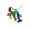
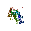
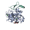
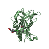
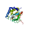
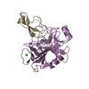
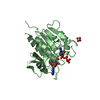
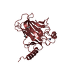
- PDB-1eug: CRYSTAL STRUCTURE OF ESCHERICHIA COLI URACIL DNA GLYCOSYLASE AND ... -
+ Open data

- Basic information
| Entry | Database: PDB / ID: 1eug | ||||||
|---|---|---|---|---|---|---|---|
| Title | CRYSTAL STRUCTURE OF ESCHERICHIA COLI URACIL DNA GLYCOSYLASE AND ITS COMPLEXES WITH URACIL AND GLYCEROL: STRUCTURE AND GLYCOSYLASE MECHANISM REVISITED | ||||||
 Components Components | PROTEIN (GLYCOSYLASE) | ||||||
 Keywords Keywords | HYDROLASE / GLYCOSYLASE | ||||||
| Function / homology |  Function and homology information Function and homology informationbase-excision repair, AP site formation via deaminated base removal / uracil-DNA glycosylase / uracil DNA N-glycosylase activity / cytoplasm Similarity search - Function | ||||||
| Biological species |  | ||||||
| Method |  X-RAY DIFFRACTION / MOLECULAR REPLACEMENT / Resolution: 1.6 Å X-RAY DIFFRACTION / MOLECULAR REPLACEMENT / Resolution: 1.6 Å | ||||||
 Authors Authors | Xiao, G. / Tordova, M. / Jagadeesh, J. / Drohat, A.C. / Stivers, J.T. / Gilliland, G.L. | ||||||
 Citation Citation | Journal: Proteins / Year: 1999 Title: Crystal structure of Escherichia coli uracil DNA glycosylase and its complexes with uracil and glycerol: structure and glycosylase mechanism revisited. Authors: Xiao, G. / Tordova, M. / Jagadeesh, J. / Drohat, A.C. / Stivers, J.T. / Gilliland, G.L. | ||||||
| History |
|
- Structure visualization
| Structure viewer | Molecule: MolmilJmol/JSmol |
|---|
- Downloads & links
-Download
| PDBx/mmCIF format | 1eug.cif.gz | 64.1 KB | Display | PDBx/mmCIF format |
|---|---|---|---|---|
| PDB format | pdb1eug.ent.gz | 45.4 KB | Display | PDB format |
| PDBx/mmJSON format | 1eug.json.gz | Tree view | PDBx/mmJSON format | |
| Others |  Other downloads Other downloads |
-Validation report
| Arichive directory | https://data.pdbj.org/pub/pdb/validation_reports/eu/1eugftp://data.pdbj.org/pub/pdb/validation_reports/eu/1eug | HTTPS FTP |
|---|
-Related structure data
| Related structure data |  2eugC 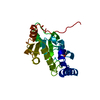 3eugC  5eugC 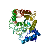 1udgS S: Starting model for refinement C: citing same article ( |
|---|---|
| Similar structure data |


-Links
 PDBj
PDBj- Assembly
| Deposited unit | 
| ||||||||
|---|---|---|---|---|---|---|---|---|---|
| 1 |
| ||||||||
| Unit cell |
|
-Components
| #1: Protein | Mass: 25706.135 Da / Num. of mol.: 1 Source method: isolated from a genetically manipulated source Source: (gene. exp.) |
|---|---|
| #2: Water | ChemComp-HOH /  Mass: 18.015 Da / Num. of mol.: 300 / Source method: isolated from a natural source / Formula: H2O Mass: 18.015 Da / Num. of mol.: 300 / Source method: isolated from a natural source / Formula: H2O |
-Experimental details
-Experiment
| Experiment | Method: X-RAY DIFFRACTION / Number of used crystals: 1 |
|---|
- Sample preparation
| Crystal | Density Matthews: 2.05 Å3/Da / Density % sol: 39.9 % | |||||||||||||||||||||||||
|---|---|---|---|---|---|---|---|---|---|---|---|---|---|---|---|---|---|---|---|---|---|---|---|---|---|---|
| Crystal grow | pH: 8.5 Details: PROTEIN CONCENTRATION 14.9 MG/ML, 0.2 M SODIUM ACETATE, 30% PEG4000, 0.1 M TRIS BUFFER, PH 8.5 USING HANGING DROP AT 293K. | |||||||||||||||||||||||||
| Crystal grow | *PLUS Temperature: 20 ℃ / Method: vapor diffusion, hanging drop | |||||||||||||||||||||||||
| Components of the solutions | *PLUS
|
-Data collection
| Diffraction | Mean temperature: 100 K |
|---|---|
| Diffraction source | Source: ROTATING ANODE / Type: BRUKER / Wavelength: 1.5418 |
| Detector | Type: BRUKER / Detector: AREA DETECTOR / Date: Aug 15, 1997 |
| Radiation | Monochromator: GRAPHITE CRYSTAL / Protocol: SINGLE WAVELENGTH / Monochromatic (M) / Laue (L): M / Scattering type: x-ray |
| Radiation wavelength | Wavelength: 1.5418 Å / Relative weight: 1 |
| Reflection | Resolution: 1.6→99 Å / Num. obs: 27664 / % possible obs: 97.9 % / Observed criterion σ(I): 1.5 / Redundancy: 4.2 % / Rmerge(I) obs: 0.095 / Net I/σ(I): 7.3 |
| Reflection shell | Resolution: 1.6→1.7 Å / Redundancy: 2.9 % / Rmerge(I) obs: 0.28 / Mean I/σ(I) obs: 3.1 / % possible all: 97.9 |
| Reflection | *PLUS Num. obs: 7194 / % possible obs: 92.2 % / Redundancy: 5.3 % / Rmerge(I) obs: 0.065 |
- Processing
| Software |
| |||||||||||||||||||||||||||||||||
|---|---|---|---|---|---|---|---|---|---|---|---|---|---|---|---|---|---|---|---|---|---|---|---|---|---|---|---|---|---|---|---|---|---|---|
| Refinement | Method to determine structure: MOLECULAR REPLACEMENT Starting model: PDB ENTRY 1UDG Resolution: 1.6→99 Å / Num. parameters: 8488 / Num. restraintsaints: 7462 / Cross valid method: FREE R / σ(F): 1 / Stereochemistry target values: ENGH AND HUBER
| |||||||||||||||||||||||||||||||||
| Solvent computation | Solvent model: MOEWS & KRETSINGER | |||||||||||||||||||||||||||||||||
| Refine analyze | Occupancy sum hydrogen: 0 / Occupancy sum non hydrogen: 2117 | |||||||||||||||||||||||||||||||||
| Refinement step | Cycle: LAST / Resolution: 1.6→99 Å
| |||||||||||||||||||||||||||||||||
| Refine LS restraints |
| |||||||||||||||||||||||||||||||||
| Software | *PLUS Name: SHELXL-97 / Classification: refinement | |||||||||||||||||||||||||||||||||
| Refinement | *PLUS Lowest resolution: 99 Å / % reflection Rfree: 5 % / Rfactor Rfree: 0.25 / Rfactor Rwork: 0.202 | |||||||||||||||||||||||||||||||||
| Solvent computation | *PLUS | |||||||||||||||||||||||||||||||||
| Displacement parameters | *PLUS | |||||||||||||||||||||||||||||||||
| Refine LS restraints | *PLUS Type: s_plane_restr / Dev ideal: 0.027 |
