 Movie
Movie Controller

[English] 日本語
 Yorodumi
- PDB-1etu: STRUCTURAL DETAILS OF THE BINDING OF GUANOSINE DIPHOSPHATE TO ELO... -
+ Open data

- Basic information
| Entry | Database: PDB / ID: 1etu | ||||||
|---|---|---|---|---|---|---|---|
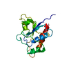
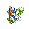
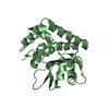
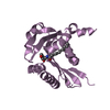
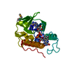
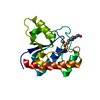
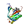
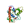
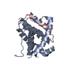
| Title | STRUCTURAL DETAILS OF THE BINDING OF GUANOSINE DIPHOSPHATE TO ELONGATION FACTOR TU FROM E. COLI AS STUDIED BY X-RAY CRYSTALLOGRAPHY | ||||||
 Components Components | ELONGATION FACTOR TU | ||||||
 Keywords Keywords | TRANSPORT AND PROTECTION PROTEIN | ||||||
| Function / homology |  Function and homology information Function and homology informationguanyl-nucleotide exchange factor complex / protein-synthesizing GTPase / guanosine tetraphosphate binding / translational elongation / translation elongation factor activity / response to antibiotic / GTPase activity / GTP binding / magnesium ion binding / RNA binding ...guanyl-nucleotide exchange factor complex / protein-synthesizing GTPase / guanosine tetraphosphate binding / translational elongation / translation elongation factor activity / response to antibiotic / GTPase activity / GTP binding / magnesium ion binding / RNA binding / plasma membrane / cytoplasm Similarity search - Function | ||||||
| Biological species |  | ||||||
| Method |  X-RAY DIFFRACTION / Resolution: 2.9 Å X-RAY DIFFRACTION / Resolution: 2.9 Å | ||||||
 Authors Authors | Clark, B.F.C. / Lacour, T.F.M. / Kjeldgaard, M. / Morikawa, K. / Nyborg, J. / Rubin, R. / Thirup, S. | ||||||
 Citation Citation | Journal: EMBO J. / Year: 1985 Title: Structural details of the binding of guanosine diphosphate to elongation factor Tu from E. coli as studied by X-ray crystallography. Authors: la Cour, T.F. / Nyborg, J. / Thirup, S. / Clark, B.F. #1: Journal: Science / Year: 1985Title: A Model for the Tertiary Structure of P21, the Product of the Ras Oncogene Authors: Mccormick, F. / Clark, B.F.C. / Lacour, T.F.M. / Kjeldgaard, M. / Norskov-Lauritsen, L. / Nyborg, J. #2: Journal: Gene Expression. The Translational Step and its ControlYear: 1984 Title: Structure of Bacterial Elongation Factor EF-TU and its Interaction with Aminoacyl-tRNA Authors: Clark, B.F.C. / Lacour, T.F.M. / Nielsen, K.M. / Nyborg, J. / Petersen, H.U. / Siboska, G.E. / Wikman, F.P. #3: Journal: FEBS Lett. / Year: 1981Title: Structural Features of the Gdp Binding Site of Elongation Factor TU from Escherichia Coli as Determined by X-Ray Diffraction Authors: Rubin, J.R. / Morikawa, K. / Nyborg, J. / Lacour, T.F.M. / Clark, B.F.C. / Miller, D.L. #4: Journal: J.Mol.Biol. / Year: 1978Title: High Resolution X-Ray Crystallographic Analysis of a Modified Form of the Elongation Factor TU(Colon) Guanosine Diphosphate Complex Authors: Morikawa, K. / Lacour, T.F.M. / Nyborg, J. / Rasmussen, K.M. / Miller, D.L. / Clark, B.F.C. | ||||||
| History |
|
- Structure visualization
| Structure viewer | Molecule: MolmilJmol/JSmol |
|---|
- Downloads & links
-Download
| PDBx/mmCIF format | 1etu.cif.gz | 51.6 KB | Display | PDBx/mmCIF format |
|---|---|---|---|---|
| PDB format | pdb1etu.ent.gz | 33 KB | Display | PDB format |
| PDBx/mmJSON format | 1etu.json.gz | Tree view | PDBx/mmJSON format | |
| Others |  Other downloads Other downloads |
-Validation report
| Arichive directory | https://data.pdbj.org/pub/pdb/validation_reports/et/1etuftp://data.pdbj.org/pub/pdb/validation_reports/et/1etu | HTTPS FTP |
|---|
-Related structure data
| Similar structure data |
|---|

-Links
 PDBj
PDBj


- Assembly
| Deposited unit | 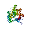
| ||||||||
|---|---|---|---|---|---|---|---|---|---|
| 1 |
| ||||||||
| Unit cell |
| ||||||||
| Atom site foot note | 1: SEE REMARK 5. |
-Components
| #1: Protein | Mass: 43209.270 Da / Num. of mol.: 1 Source method: isolated from a genetically manipulated source Source: (gene. exp.) |
|---|---|
| #2: Chemical | ChemComp-MG /   Mass: 24.305 Da / Num. of mol.: 1 / Source method: obtained synthetically / Formula: Mg Mass: 24.305 Da / Num. of mol.: 1 / Source method: obtained synthetically / Formula: Mg |
| #3: Chemical | ChemComp-GDP /   Type: RNA linking / Mass: 443.201 Da / Num. of mol.: 1 / Source method: obtained synthetically / Formula: C10H15N5O11P2 / Comment: GDP, energy-carrying molecule*YM Type: RNA linking / Mass: 443.201 Da / Num. of mol.: 1 / Source method: obtained synthetically / Formula: C10H15N5O11P2 / Comment: GDP, energy-carrying molecule*YM |
| Has protein modification | N |
| Sequence details | EF-TU IS CODED FOR BY TWO DIFFERENT GENES. THE SEQUENCE STRUCTURE ANALYSIS CARRIED OUT ON THIS ...EF-TU IS CODED FOR BY TWO DIFFERENT GENES. THE SEQUENCE STRUCTURE ANALYSIS CARRIED OUT ON THIS MIXTURE SHOWS THAT THE C-TERMINAL RESIDUE OCCURS AS GLY/SER IN THE RATIO OF 3/1. THIS RESIDUE IS IDENTIFIED |
-Experimental details
-Experiment
| Experiment | Method: X-RAY DIFFRACTION |
|---|
- Sample preparation
| Crystal | Density Matthews: 4.6 Å3/Da / Density % sol: 73.26 % | ||||||||||||||||||||||||||||||||||||||||||
|---|---|---|---|---|---|---|---|---|---|---|---|---|---|---|---|---|---|---|---|---|---|---|---|---|---|---|---|---|---|---|---|---|---|---|---|---|---|---|---|---|---|---|---|
| Crystal grow | *PLUS pH: 7 / Method: vapor diffusion / Details: referred to J.Mol.Biol. 125.325-338 | ||||||||||||||||||||||||||||||||||||||||||
| Components of the solutions | *PLUS
|
- Processing
| Refinement | Highest resolution: 2.9 Å Details: ATOMS WITH AN OCCUPANCY OF 0.0 ARE POORLY DEFINED IN THE DENSITY. | ||||||||||||
|---|---|---|---|---|---|---|---|---|---|---|---|---|---|
| Refinement step | Cycle: LAST / Highest resolution: 2.9 Å
|
