 Movie
Movie Controller

[English] 日本語
 Yorodumi
- PDB-1et0: CRYSTAL STRUCTURE OF AMINODEOXYCHORISMATE LYASE FROM ESCHERICHIA COLI -
+ Open data

- Basic information
| Entry | Database: PDB / ID: 1et0 | ||||||
|---|---|---|---|---|---|---|---|
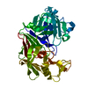
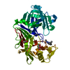
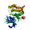
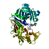
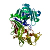
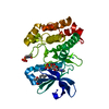
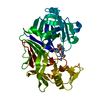
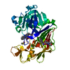
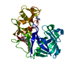
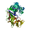
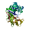
| Title | CRYSTAL STRUCTURE OF AMINODEOXYCHORISMATE LYASE FROM ESCHERICHIA COLI | ||||||
 Components Components | 4-AMINO-4-DEOXYCHORISMATE LYASE | ||||||
 Keywords Keywords | LYASE / PSEUDO BETA BARREL | ||||||
| Function / homology |  Function and homology information Function and homology information4-amino-4-deoxychorismate lyase activity / aminodeoxychorismate lyase / 4-aminobenzoate biosynthetic process / folic acid biosynthetic process / tetrahydrofolate biosynthetic process / pyridoxal phosphate binding / cytosol Similarity search - Function | ||||||
| Biological species |  | ||||||
| Method |  X-RAY DIFFRACTION / SYNCHROTRON / Resolution: 2.2 Å X-RAY DIFFRACTION / SYNCHROTRON / Resolution: 2.2 Å | ||||||
 Authors Authors | Nakai, T. / Mizutani, H. / Miyahara, I. / Hirotsu, K. / Takeda, S. / Jhee, K.H. / Yoshimura, T. / Esaki, N. | ||||||
 Citation Citation | Journal: J.Biochem.(Tokyo) / Year: 2000 Title: Three-dimensional structure of 4-amino-4-deoxychorismate lyase from Escherichia coli. Authors: Nakai, T. / Mizutani, H. / Miyahara, I. / Hirotsu, K. / Takeda, S. / Jhee, K.H. / Yoshimura, T. / Esaki, N. #1: Journal: J.Bacteriol. / Year: 1992Title: Characterization and Sequence of Escherichia coli pabC, the Gene Encoding Aminodeoxychorismate Lyase, a Pyridoxal Phosphate-Containing Enzyme Authors: Green, J.M. / Merkel, W.K. / Nichols, B.P. | ||||||
| History |
|
- Structure visualization
| Structure viewer | Molecule: MolmilJmol/JSmol |
|---|
- Downloads & links
-Download
| PDBx/mmCIF format | 1et0.cif.gz | 65 KB | Display | PDBx/mmCIF format |
|---|---|---|---|---|
| PDB format | pdb1et0.ent.gz | 46.7 KB | Display | PDB format |
| PDBx/mmJSON format | 1et0.json.gz | Tree view | PDBx/mmJSON format | |
| Others |  Other downloads Other downloads |
-Validation report
| Summary document | 1et0_validation.pdf.gz | 387.2 KB | Display | wwPDB validaton report |
|---|---|---|---|---|
| Full document | 1et0_full_validation.pdf.gz | 390.7 KB | Display | |
| Data in XML | 1et0_validation.xml.gz | 7.1 KB | Display | |
| Data in CIF | 1et0_validation.cif.gz | 11 KB | Display | |
| Arichive directory | https://data.pdbj.org/pub/pdb/validation_reports/et/1et0ftp://data.pdbj.org/pub/pdb/validation_reports/et/1et0 | HTTPS FTP |
-Related structure data
| Similar structure data |
|---|


-Links
 PDBj
PDBj
- Assembly
| Deposited unit | 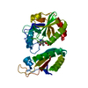
| ||||||||
|---|---|---|---|---|---|---|---|---|---|
| 1 |
| ||||||||
| 2 | 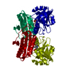
| ||||||||
| Unit cell |
| ||||||||
| Details | The biological assembly is a dimer constructed from chain A a symmetry partner generated by the two-fold. |
-Components
| #1: Protein | Mass: 29745.031 Da / Num. of mol.: 1 Source method: isolated from a genetically manipulated source Source: (gene. exp.) |
|---|---|
| #2: Chemical | ChemComp-PLP /   Mass: 247.142 Da / Num. of mol.: 1 / Source method: obtained synthetically / Formula: C8H10NO6P Mass: 247.142 Da / Num. of mol.: 1 / Source method: obtained synthetically / Formula: C8H10NO6P |
| #3: Water | ChemComp-HOH /  Mass: 18.015 Da / Num. of mol.: 126 / Source method: isolated from a natural source / Formula: H2O Mass: 18.015 Da / Num. of mol.: 126 / Source method: isolated from a natural source / Formula: H2O |
| Has protein modification | N |
-Experimental details
-Experiment
| Experiment | Method: X-RAY DIFFRACTION / Number of used crystals: 1 |
|---|
- Sample preparation
| Crystal | Density Matthews: 2.11 Å3/Da / Density % sol: 41.61 % | |||||||||||||||||||||||||||||||||||
|---|---|---|---|---|---|---|---|---|---|---|---|---|---|---|---|---|---|---|---|---|---|---|---|---|---|---|---|---|---|---|---|---|---|---|---|---|
| Crystal grow | Temperature: 293 K / Method: vapor diffusion, hanging drop / pH: 6.5 Details: PEG 400, 2-propanol, sodium citrate, pH 6.5, VAPOR DIFFUSION, HANGING DROP, temperature 293K | |||||||||||||||||||||||||||||||||||
| Crystal grow | *PLUS Details: drop consists of equal volume of protein and reservoir solutions | |||||||||||||||||||||||||||||||||||
| Components of the solutions | *PLUS
|
-Data collection
| Diffraction | Mean temperature: 287 K |
|---|---|
| Diffraction source | Source: SYNCHROTRON / Site: Photon Factory  / Beamline: BL-6A / Wavelength: 1 / Beamline: BL-6A / Wavelength: 1 |
| Detector | Type: FUJI / Detector: IMAGE PLATE / Date: Jan 20, 1999 |
| Radiation | Protocol: SINGLE WAVELENGTH / Monochromatic (M) / Laue (L): M / Scattering type: x-ray |
| Radiation wavelength | Wavelength: 1 Å / Relative weight: 1 |
| Reflection | Resolution: 2.2→50 Å / Num. all: 13365 / Num. obs: 12330 / % possible obs: 92.2 % / Observed criterion σ(F): 0 / Observed criterion σ(I): 0 / Redundancy: 7.14 % / Biso Wilson estimate: 21.4 Å2 / Rmerge(I) obs: 0.072 / Net I/σ(I): 30.9 |
| Reflection shell | Resolution: 2.2→2.28 Å / Redundancy: 5.1 % / Rmerge(I) obs: 0.135 / Num. unique all: 1110 / % possible all: 85 |
| Reflection | *PLUS Num. measured all: 88073 |
| Reflection shell | *PLUS % possible obs: 85 % |
- Processing
| Software |
| |||||||||||||||||||||||||
|---|---|---|---|---|---|---|---|---|---|---|---|---|---|---|---|---|---|---|---|---|---|---|---|---|---|---|
| Refinement | Resolution: 2.2→8 Å / σ(F): 2 / σ(I): 0 / Stereochemistry target values: Engh & Huber
| |||||||||||||||||||||||||
| Refinement step | Cycle: LAST / Resolution: 2.2→8 Å
| |||||||||||||||||||||||||
| Refine LS restraints |
|
