 Movie
Movie Controller

[English] 日本語
 Yorodumi
- PDB-1eq2: THE CRYSTAL STRUCTURE OF ADP-L-GLYCERO-D-MANNOHEPTOSE 6-EPIMERASE -
+ Open data

- Basic information
| Entry | Database: PDB / ID: 1eq2 | ||||||
|---|---|---|---|---|---|---|---|
| Title | THE CRYSTAL STRUCTURE OF ADP-L-GLYCERO-D-MANNOHEPTOSE 6-EPIMERASE | ||||||
 Components Components | ADP-L-GLYCERO-D-MANNOHEPTOSE 6-EPIMERASE | ||||||
 Keywords Keywords | ISOMERASE / N-terminal domain Rossmann fold / C-terminal mixed alpha/beta domain / Short-chain dehydrogenase/reductase fold | ||||||
| Function / homology |  Function and homology information Function and homology informationADP-glyceromanno-heptose 6-epimerase / ADP-glyceromanno-heptose 6-epimerase activity / ADP-L-glycero-beta-D-manno-heptose biosynthetic process / lipopolysaccharide core region biosynthetic process / NADP+ binding / membrane / cytosol Similarity search - Function | ||||||
| Biological species |  | ||||||
| Method |  X-RAY DIFFRACTION / SYNCHROTRON / Resolution: 2 Å X-RAY DIFFRACTION / SYNCHROTRON / Resolution: 2 Å | ||||||
 Authors Authors | Deacon, A.M. / Ni, Y.S. / Coleman Jr., W.G. / Ealick, S.E. | ||||||
 Citation Citation | Journal: Structure Fold.Des. / Year: 2000 Title: The crystal structure of ADP-L-glycero-D-mannoheptose 6-epimerase: catalysis with a twist. Authors: Deacon, A.M. / Ni, Y.S. / Coleman Jr., W.G. / Ealick, S.E. #1: Journal: Acta Crystallogr.,Sect.D / Year: 1999Title: Crystallization and Preliminary X-ray Diffraction Studies of the Lipopolysaccharide Core Biosynthetic Enzyme ADP-L-glycero-D-mannoheptose 6-epimerase from Escherichia coli K-12 Authors: Ding, L. / Zhang, Y. / Deacon, A.M. / Ealick, S.E. / Ni, Y. / Sun, P. / Coleman Jr., W.G. | ||||||
| History |
|
- Structure visualization
| Structure viewer | Molecule: MolmilJmol/JSmol |
|---|
- Downloads & links
-Download
| PDBx/mmCIF format | 1eq2.cif.gz | 622.3 KB | Display | PDBx/mmCIF format |
|---|---|---|---|---|
| PDB format | pdb1eq2.ent.gz | 513.1 KB | Display | PDB format |
| PDBx/mmJSON format | 1eq2.json.gz | Tree view | PDBx/mmJSON format | |
| Others |  Other downloads Other downloads |
-Validation report
| Summary document | 1eq2_validation.pdf.gz | 1.6 MB | Display | wwPDB validaton report |
|---|---|---|---|---|
| Full document | 1eq2_full_validation.pdf.gz | 1.7 MB | Display | |
| Data in XML | 1eq2_validation.xml.gz | 70.7 KB | Display | |
| Data in CIF | 1eq2_validation.cif.gz | 98.3 KB | Display | |
| Arichive directory | https://data.pdbj.org/pub/pdb/validation_reports/eq/1eq2ftp://data.pdbj.org/pub/pdb/validation_reports/eq/1eq2 | HTTPS FTP |
-Related structure data
| Similar structure data |
|---|

-Links
 PDBj
PDBj- Assembly
| Deposited unit | 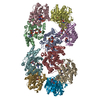
| ||||||||
|---|---|---|---|---|---|---|---|---|---|
| 1 | 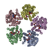
| ||||||||
| 2 | 
| ||||||||
| Unit cell |
| ||||||||
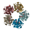
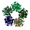
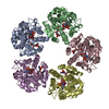
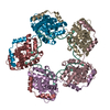
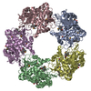
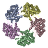
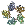
| Details | The biological assembly is a pentamer. There are two pentamers in the asymmetric unit, each with a local non-crystallographic five-fold axis parallel to the crystallographic two-fold screw axis. |
-Components
| #1: Protein | Mass: 34943.031 Da / Num. of mol.: 10 Source method: isolated from a genetically manipulated source Source: (gene. exp.) References: UniProt: P67910, ADP-glyceromanno-heptose 6-epimerase #2: Chemical | ChemComp-NAP /   Mass: 743.405 Da / Num. of mol.: 10 / Source method: obtained synthetically / Formula: C21H28N7O17P3 / Details: Sigma Mass: 743.405 Da / Num. of mol.: 10 / Source method: obtained synthetically / Formula: C21H28N7O17P3 / Details: Sigma#3: Chemical | ChemComp-ADQ / 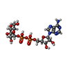  Mass: 589.342 Da / Num. of mol.: 10 / Source method: obtained synthetically / Formula: C16H25N5O15P2 / Details: Sigma Mass: 589.342 Da / Num. of mol.: 10 / Source method: obtained synthetically / Formula: C16H25N5O15P2 / Details: Sigma#4: Water | ChemComp-HOH / |  Mass: 18.015 Da / Num. of mol.: 1056 / Source method: isolated from a natural source / Formula: H2O Mass: 18.015 Da / Num. of mol.: 1056 / Source method: isolated from a natural source / Formula: H2OHas protein modification | Y | |
|---|
-Experimental details
-Experiment
| Experiment | Method: X-RAY DIFFRACTION / Number of used crystals: 1 |
|---|
- Sample preparation
| Crystal | Density Matthews: 2.83 Å3/Da / Density % sol: 56.61 % | ||||||||||||||||||||||||||||||||||||||||||||||||||||||||
|---|---|---|---|---|---|---|---|---|---|---|---|---|---|---|---|---|---|---|---|---|---|---|---|---|---|---|---|---|---|---|---|---|---|---|---|---|---|---|---|---|---|---|---|---|---|---|---|---|---|---|---|---|---|---|---|---|---|
| Crystal grow | Temperature: 295 K / Method: vapor diffusion, hanging drop / pH: 7.5 Details: 2.0M ammonium sulphate, 2% PEG400, 0.1M HEPES buffer, 1mM ADP-glucose, 20nM spermidine, pH 7.5, VAPOR DIFFUSION, HANGING DROP, temperature 295K | ||||||||||||||||||||||||||||||||||||||||||||||||||||||||
| Crystal grow | *PLUS | ||||||||||||||||||||||||||||||||||||||||||||||||||||||||
| Components of the solutions | *PLUS
|
-Data collection
| Diffraction | Mean temperature: 100 K |
|---|---|
| Diffraction source | Source: SYNCHROTRON / Site: CHESS  / Beamline: F1 / Wavelength: 0.919 / Beamline: F1 / Wavelength: 0.919 |
| Detector | Type: ADSC QUANTUM 4 / Detector: CCD / Date: Dec 22, 1998 |
| Radiation | Protocol: SINGLE WAVELENGTH / Monochromatic (M) / Laue (L): M / Scattering type: x-ray |
| Radiation wavelength | Wavelength: 0.919 Å / Relative weight: 1 |
| Reflection | Resolution: 2→25 Å / Num. all: 242386 / Num. obs: 242386 / % possible obs: 93.3 % / Observed criterion σ(F): 0 / Observed criterion σ(I): 0 / Redundancy: 2.4 % / Biso Wilson estimate: 25.4 Å2 / Rmerge(I) obs: 0.066 / Net I/σ(I): 11.7 |
| Reflection shell | Resolution: 2→2.11 Å / Redundancy: 3 % / Rmerge(I) obs: 0.218 / Num. unique all: 239829 / % possible all: 78.6 |
| Reflection | *PLUS Num. measured all: 573756 |
| Reflection shell | *PLUS % possible obs: 78.6 % / Mean I/σ(I) obs: 3 |
- Processing
| Software |
| |||||||||||||||||||||||||
|---|---|---|---|---|---|---|---|---|---|---|---|---|---|---|---|---|---|---|---|---|---|---|---|---|---|---|
| Refinement | Resolution: 2→20 Å / σ(F): 2 / σ(I): 0 / Stereochemistry target values: Engh & Huber
| |||||||||||||||||||||||||
| Refinement step | Cycle: LAST / Resolution: 2→20 Å
| |||||||||||||||||||||||||
| Refine LS restraints |
| |||||||||||||||||||||||||
| Software | *PLUS Name: X-PLOR / Version: 3.843 / Classification: refinement | |||||||||||||||||||||||||
| Refinement | *PLUS Highest resolution: 2 Å / Lowest resolution: 20 Å / σ(F): 2 | |||||||||||||||||||||||||
| Solvent computation | *PLUS | |||||||||||||||||||||||||
| Displacement parameters | *PLUS | |||||||||||||||||||||||||
| Refine LS restraints | *PLUS Type: x_angle_deg / Dev ideal: 1.5 |
