 Movie
Movie Controller

[English] 日本語
 Yorodumi
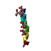
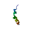
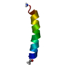
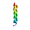
- PDB-1emz: SOLUTION STRUCTURE OF FRAGMENT (350-370) OF THE TRANSMEMBRANE DOM... -
+ Open data

- Basic information
| Entry | Database: PDB / ID: 1emz | ||||||
|---|---|---|---|---|---|---|---|
| Title | SOLUTION STRUCTURE OF FRAGMENT (350-370) OF THE TRANSMEMBRANE DOMAIN OF HEPATITIS C ENVELOPE GLYCOPROTEIN E1 | ||||||
 Components Components | ENVELOPE GLYCOPROTEIN E1 | ||||||
 Keywords Keywords | VIRAL PROTEIN / transmembrane domain / envelope protein E1 / hepatitis C virus | ||||||
| Function / homology |  Function and homology information Function and homology informationhost cell lipid droplet / host cell endoplasmic reticulum membrane / fusion of virus membrane with host endosome membrane / viral envelope / symbiont entry into host cell / virion attachment to host cell / virion membrane Similarity search - Function | ||||||
| Method | SOLUTION NMR / distance geometry, simulated annealing, molecular dynamic, energy minimization | ||||||
 Authors Authors | Op De Beeck, A. / Montserret, R. / Duvet, S. / Cocquerel, L. / Cacan, R. / Barberot, B. / Le Maire, M. / Penin, F. / Dubuisson, J. | ||||||
 Citation Citation | Journal: J.Biol.Chem. / Year: 2000 Title: The transmembrane domains of hepatitis C virus envelope glycoproteins E1 and E2 play a major role in heterodimerization. Authors: Op De Beeck, A. / Montserret, R. / Duvet, S. / Cocquerel, L. / Cacan, R. / Barberot, B. / Le Maire, M. / Penin, F. / Dubuisson, J. | ||||||
| History |
|
- Structure visualization
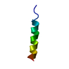
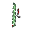
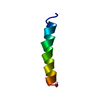
| Structure viewer | Molecule: MolmilJmol/JSmol |
|---|
- Downloads & links
-Download
| PDBx/mmCIF format | 1emz.cif.gz | 13.7 KB | Display | PDBx/mmCIF format |
|---|---|---|---|---|
| PDB format | pdb1emz.ent.gz | 7.3 KB | Display | PDB format |
| PDBx/mmJSON format | 1emz.json.gz | Tree view | PDBx/mmJSON format | |
| Others |  Other downloads Other downloads |
-Validation report
| Summary document | 1emz_validation.pdf.gz | 247.7 KB | Display | wwPDB validaton report |
|---|---|---|---|---|
| Full document | 1emz_full_validation.pdf.gz | 247.5 KB | Display | |
| Data in XML | 1emz_validation.xml.gz | 1.6 KB | Display | |
| Data in CIF | 1emz_validation.cif.gz | 1.6 KB | Display | |
| Arichive directory | https://data.pdbj.org/pub/pdb/validation_reports/em/1emzftp://data.pdbj.org/pub/pdb/validation_reports/em/1emz | HTTPS FTP |
-Related structure data
| Similar structure data |
|---|


-Links
 PDBj
PDBj
- Assembly
| Deposited unit | 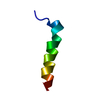
| |||||||||
|---|---|---|---|---|---|---|---|---|---|---|
| 1 |
| |||||||||
| NMR ensembles |
|
-Components
| #1: Protein/peptide | Mass: 2237.581 Da / Num. of mol.: 1 / Fragment: TRANSMEMBRANE DOMAIN (RESIDUES 350-370) / Source method: obtained synthetically Details: This peptide was chemically synthesized. The sequence of this peptide is naturally found in hepatitis C virus. References: UniProt: Q9Q3N3 |
|---|
-Experimental details
-Experiment
| Experiment | Method: SOLUTION NMR | ||||||||||||||||
|---|---|---|---|---|---|---|---|---|---|---|---|---|---|---|---|---|---|
| NMR experiment |
| ||||||||||||||||
| NMR details | Text: sodium 2,2 dimethyl-2-silapentane-5-sulfonate (DSS) in the internal nmr reference |
- Sample preparation
| Details | Contents: 4 mM E1(350-370), 50% H2O, 50% D2 trifluoroethanol (v/v) Solvent system: 50% H2O, 50% D2 trifluoroethanol (v/v) |
|---|---|
| Sample conditions | pH: 5.7 / Pressure: ambient / Temperature: 293 K |
| Crystal grow | *PLUS Method: other / Details: NMR |
-NMR measurement
| NMR spectrometer | Type: Varian UNITYPLUS / Manufacturer: Varian / Model: UNITYPLUS / Field strength: 500 MHz |
|---|
- Processing
| NMR software |
| ||||||||||||||||
|---|---|---|---|---|---|---|---|---|---|---|---|---|---|---|---|---|---|
| Refinement | Method: distance geometry, simulated annealing, molecular dynamic, energy minimization Software ordinal: 1 Details: the structure is based on 337 Noe derived distance constraints | ||||||||||||||||
| NMR representative | Selection criteria: closest to the average | ||||||||||||||||
| NMR ensemble | Conformer selection criteria: back calculated data agree with experimental NOESY spectrum,structures with the least restraint violations,structures with the lowest energy Conformers calculated total number: 50 / Conformers submitted total number: 1 |
 X-PLOR