 Movie
Movie Controller

[English] 日本語
 Yorodumi
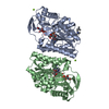
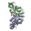
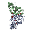
- PDB-1ek5: STRUCTURE OF HUMAN UDP-GALACTOSE 4-EPIMERASE IN COMPLEX WITH NAD+ -
+ Open data

- Basic information
| Entry | Database: PDB / ID: 1ek5 | ||||||
|---|---|---|---|---|---|---|---|
| Title | STRUCTURE OF HUMAN UDP-GALACTOSE 4-EPIMERASE IN COMPLEX WITH NAD+ | ||||||
 Components Components | UDP-GALACTOSE 4-EPIMERASE | ||||||
 Keywords Keywords | ISOMERASE / Short-Chain Dehydrogenase / Epimerase / Galactosemia | ||||||
| Function / homology |  Function and homology information Function and homology informationDefective GALE causes EDG / UDP-N-acetylglucosamine 4-epimerase / UDP-N-acetylglucosamine 4-epimerase activity / galactose catabolic process / UDP-glucose 4-epimerase / UDP-glucose 4-epimerase activity / beta-D-galactose catabolic process via UDP-galactose, Leloir pathway / Galactose catabolism / protein homodimerization activity / identical protein binding / cytosol Similarity search - Function | ||||||
| Biological species |  Homo sapiens (human) Homo sapiens (human) | ||||||
| Method |  X-RAY DIFFRACTION / Resolution: 1.8 Å X-RAY DIFFRACTION / Resolution: 1.8 Å | ||||||
 Authors Authors | Thoden, J.B. / Wohlers, T.M. / Fridovich-Keil, J.L. / Holden, H.M. | ||||||
 Citation Citation | Journal: Biochemistry / Year: 2000 Title: Crystallographic evidence for Tyr 157 functioning as the active site base in human UDP-galactose 4-epimerase. Authors: Thoden, J.B. / Wohlers, T.M. / Fridovich-Keil, J.L. / Holden, H.M. | ||||||
| History |
|
- Structure visualization
| Structure viewer | Molecule: MolmilJmol/JSmol |
|---|
- Downloads & links
-Download
| PDBx/mmCIF format | 1ek5.cif.gz | 84.9 KB | Display | PDBx/mmCIF format |
|---|---|---|---|---|
| PDB format | pdb1ek5.ent.gz | 63.5 KB | Display | PDB format |
| PDBx/mmJSON format | 1ek5.json.gz | Tree view | PDBx/mmJSON format | |
| Others |  Other downloads Other downloads |
-Validation report
| Arichive directory | https://data.pdbj.org/pub/pdb/validation_reports/ek/1ek5ftp://data.pdbj.org/pub/pdb/validation_reports/ek/1ek5 | HTTPS FTP |
|---|
-Related structure data


-Links
 PDBj
PDBj- Assembly
| Deposited unit | 
| ||||||||
|---|---|---|---|---|---|---|---|---|---|
| 1 | 
| ||||||||
| Unit cell |
| ||||||||
| Details | The biological assembly is a dimer constructed from a symmetry partner generated by the two-fold. |
-Components
| #1: Protein | Mass: 38324.605 Da / Num. of mol.: 1 Source method: isolated from a genetically manipulated source Source: (gene. exp.) Homo sapiens (human) / Tissue: FORESKIN / Production host:  |
|---|---|
| #2: Chemical | ChemComp-NAD /   Mass: 663.425 Da / Num. of mol.: 1 / Source method: obtained synthetically / Formula: C21H27N7O14P2 / Comment: NAD*YM Mass: 663.425 Da / Num. of mol.: 1 / Source method: obtained synthetically / Formula: C21H27N7O14P2 / Comment: NAD*YM |
| #3: Water | ChemComp-HOH /  Mass: 18.015 Da / Num. of mol.: 196 / Source method: isolated from a natural source / Formula: H2O Mass: 18.015 Da / Num. of mol.: 196 / Source method: isolated from a natural source / Formula: H2O |
-Experimental details
-Experiment
| Experiment | Method: X-RAY DIFFRACTION / Number of used crystals: 1 |
|---|
- Sample preparation
| Crystal | Density Matthews: 2.49 Å3/Da / Density % sol: 50.59 % | ||||||||||||||||||||||||||||||||||||||||||
|---|---|---|---|---|---|---|---|---|---|---|---|---|---|---|---|---|---|---|---|---|---|---|---|---|---|---|---|---|---|---|---|---|---|---|---|---|---|---|---|---|---|---|---|
| Crystal grow | Temperature: 298 K / Method: vapor diffusion, hanging drop / pH: 7.5 Details: PEG-8000, KCl, HEPPS, pH 7.5, VAPOR DIFFUSION, HANGING DROP, temperature 298.0K | ||||||||||||||||||||||||||||||||||||||||||
| Crystal grow | *PLUS | ||||||||||||||||||||||||||||||||||||||||||
| Components of the solutions | *PLUS
|
-Data collection
| Diffraction | Mean temperature: 273 K |
|---|---|
| Diffraction source | Source: ROTATING ANODE / Type: RIGAKU RU200 / Wavelength: 1.5418 |
| Detector | Type: SIEMENS HI-STAR / Detector: AREA DETECTOR / Date: May 27, 1999 |
| Radiation | Protocol: SINGLE WAVELENGTH / Monochromatic (M) / Laue (L): M / Scattering type: x-ray |
| Radiation wavelength | Wavelength: 1.5418 Å / Relative weight: 1 |
| Reflection | Resolution: 1.8→30 Å / Num. all: 34184 / Num. obs: 34184 / % possible obs: 95.2 % / Observed criterion σ(F): 0 / Observed criterion σ(I): 0 / Redundancy: 2.9 % / Rmerge(I) obs: 0.059 / Net I/σ(I): 16.5 |
| Reflection shell | Resolution: 1.8→1.88 Å / Redundancy: 1.8 % / Rmerge(I) obs: 0.258 / Num. unique all: 3761 / % possible all: 87 |
| Reflection | *PLUS |
| Reflection shell | *PLUS % possible obs: 87 % / Num. unique obs: 3761 / Mean I/σ(I) obs: 3 |
- Processing
| Software |
| |||||||||||||||||||||||||
|---|---|---|---|---|---|---|---|---|---|---|---|---|---|---|---|---|---|---|---|---|---|---|---|---|---|---|
| Refinement | Resolution: 1.8→30 Å / σ(F): 0 / σ(I): 0 / Stereochemistry target values: Engh & Huber
| |||||||||||||||||||||||||
| Refinement step | Cycle: LAST / Resolution: 1.8→30 Å
| |||||||||||||||||||||||||
| Refine LS restraints |
| |||||||||||||||||||||||||
| Software | *PLUS Name: TNT / Version: 5E / Classification: refinement | |||||||||||||||||||||||||
| Refinement | *PLUS σ(F): 0 / Rfactor all: 0.191 / Rfactor Rwork: 0.19 | |||||||||||||||||||||||||
| Solvent computation | *PLUS | |||||||||||||||||||||||||
| Displacement parameters | *PLUS | |||||||||||||||||||||||||
| Refine LS restraints | *PLUS
|
