 Movie
Movie Controller


+ Open data

- Basic information
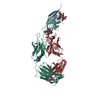
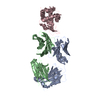
| Entry | Database: PDB / ID: 1egj | ||||||
|---|---|---|---|---|---|---|---|
| Title | DOMAIN 4 OF THE BETA COMMON CHAIN IN COMPLEX WITH AN ANTIBODY | ||||||
 Components Components |
| ||||||
 Keywords Keywords | IMMUNE SYSTEM / cytokine receptor complexed to an antibody | ||||||
| Function / homology |  Function and homology information Function and homology informationDefective CSF2RB causes SMDP5 / Defective CSF2RA causes SMDP4 / positive regulation of type IIa hypersensitivity / positive regulation of B cell activation / humoral immune response mediated by circulating immunoglobulin / phagocytosis, recognition / granulocyte macrophage colony-stimulating factor receptor complex / granulocyte-macrophage colony-stimulating factor signaling pathway / early endosome to late endosome transport / respiratory gaseous exchange by respiratory system ...Defective CSF2RB causes SMDP5 / Defective CSF2RA causes SMDP4 / positive regulation of type IIa hypersensitivity / positive regulation of B cell activation / humoral immune response mediated by circulating immunoglobulin / phagocytosis, recognition / granulocyte macrophage colony-stimulating factor receptor complex / granulocyte-macrophage colony-stimulating factor signaling pathway / early endosome to late endosome transport / respiratory gaseous exchange by respiratory system / interleukin-5-mediated signaling pathway / positive regulation of leukocyte proliferation / positive regulation of type I hypersensitivity / interleukin-3-mediated signaling pathway / cellular response to interleukin-3 / Surfactant metabolism / antibody-dependent cellular cytotoxicity / Fc-gamma receptor I complex binding / cytokine receptor activity / immunoglobulin complex, circulating / phagocytosis, engulfment / immunoglobulin receptor binding / antigen processing and presentation / IgG immunoglobulin complex / endosome to lysosome transport / immunoglobulin mediated immune response / regulation of proteolysis / Interleukin-3, Interleukin-5 and GM-CSF signaling / complement activation, classical pathway / Interleukin receptor SHC signaling / immunoglobulin complex / positive regulation of endocytosis / antigen binding / cell surface receptor signaling pathway via JAK-STAT / coreceptor activity / multivesicular body / positive regulation of phagocytosis / response to bacterium / positive regulation of immune response / antibacterial humoral response / signaling receptor activity / RAF/MAP kinase cascade / response to lipopolysaccharide / adaptive immune response / immune response / external side of plasma membrane / signal transduction / : / extracellular region / plasma membrane Similarity search - Function | ||||||
| Biological species |  Homo sapiens (human) Homo sapiens (human) | ||||||
| Method |  X-RAY DIFFRACTION / SYNCHROTRON / Resolution: 2.8 Å X-RAY DIFFRACTION / SYNCHROTRON / Resolution: 2.8 Å | ||||||
 Authors Authors | Rossjohn, J. / McKinstry, W.J. / Woodcock, J.M. / McClure, B.J. / Hercus, T.R. / Parker, M.W. / Lopez, A.F. / Bagley, C.J. | ||||||
 Citation Citation | Journal: Blood / Year: 2000 Title: Structure of the activation domain of the GM-CSF/IL-3/IL-5 receptor common beta-chain bound to an antagonist. Authors: Rossjohn, J. / McKinstry, W.J. / Woodcock, J.M. / McClure, B.J. / Hercus, T.R. / Parker, M.W. / Lopez, A.F. / Bagley, C.J. | ||||||
| History |
|
- Structure visualization
| Structure viewer | Molecule: MolmilJmol/JSmol |
|---|
- Downloads & links
-Download
| PDBx/mmCIF format | 1egj.cif.gz | 120.6 KB | Display | PDBx/mmCIF format |
|---|---|---|---|---|
| PDB format | pdb1egj.ent.gz | 92.1 KB | Display | PDB format |
| PDBx/mmJSON format | 1egj.json.gz | Tree view | PDBx/mmJSON format | |
| Others |  Other downloads Other downloads |
-Validation report
| Arichive directory | https://data.pdbj.org/pub/pdb/validation_reports/eg/1egjftp://data.pdbj.org/pub/pdb/validation_reports/eg/1egj | HTTPS FTP |
|---|
-Related structure data
| Similar structure data |
|---|


-Links
 PDBj
PDBj


- Assembly
| Deposited unit | 
| ||||||||
|---|---|---|---|---|---|---|---|---|---|
| 1 |
| ||||||||
| Unit cell |
|
-Components
| #1: Protein | Mass: 11991.282 Da / Num. of mol.: 1 / Fragment: DOMAIN 4 Source method: isolated from a genetically manipulated source Source: (gene. exp.) Homo sapiens (human) / Plasmid: PCDNA3 (INVITROGEN) / Production host:  |
|---|---|
| #2: Antibody | Mass: 23589.877 Da / Num. of mol.: 1 / Source method: isolated from a natural source / Details: MONOCLONAL ANTIBODY / Source: (natural) |
| #3: Antibody | Mass: 23380.188 Da / Num. of mol.: 1 / Source method: isolated from a natural source / Details: MONOCLONAL ANTIBODY / Source: (natural) |
| #4: Sugar | ChemComp-NAG /   Type: D-saccharide, beta linking / Mass: 221.208 Da / Num. of mol.: 1 Type: D-saccharide, beta linking / Mass: 221.208 Da / Num. of mol.: 1Source method: isolated from a genetically manipulated source Formula: C8H15NO6 |
| #5: Water | ChemComp-HOH /  Mass: 18.015 Da / Num. of mol.: 144 / Source method: isolated from a natural source / Formula: H2O Mass: 18.015 Da / Num. of mol.: 144 / Source method: isolated from a natural source / Formula: H2O |
| Has protein modification | Y |
-Experimental details
-Experiment
| Experiment | Method: X-RAY DIFFRACTION / Number of used crystals: 1 |
|---|
- Sample preparation
| Crystal | Density Matthews: 3.78 Å3/Da / Density % sol: 67.47 % | ||||||||||||||||||||||||||||||
|---|---|---|---|---|---|---|---|---|---|---|---|---|---|---|---|---|---|---|---|---|---|---|---|---|---|---|---|---|---|---|---|
| Crystal grow | Temperature: 295 K / Method: vapor diffusion, hanging drop / pH: 5.5 Details: 12% peg 4000, pH 5.5, VAPOR DIFFUSION, HANGING DROP, temperature 22K | ||||||||||||||||||||||||||||||
| Crystal grow | *PLUS | ||||||||||||||||||||||||||||||
| Components of the solutions | *PLUS
|
-Data collection
| Diffraction | Mean temperature: 100 K |
|---|---|
| Diffraction source | Source: SYNCHROTRON / Site: APS  / Beamline: 14-BM-C / Wavelength: 1 / Beamline: 14-BM-C / Wavelength: 1 |
| Detector | Type: OTHER / Detector: CCD / Date: May 1, 1999 |
| Radiation | Protocol: SINGLE WAVELENGTH / Monochromatic (M) / Laue (L): M / Scattering type: x-ray |
| Radiation wavelength | Wavelength: 1 Å / Relative weight: 1 |
| Reflection | Resolution: 2.8→30 Å / Num. all: 21211 / Num. obs: 21211 / % possible obs: 88.1 % / Observed criterion σ(F): 0 / Observed criterion σ(I): 0 / Redundancy: 2.8 % / Biso Wilson estimate: 85.6 Å2 / Rmerge(I) obs: 0.098 / Net I/σ(I): 11.4 |
| Reflection shell | Resolution: 2.8→2.9 Å / Redundancy: 2 % / Rmerge(I) obs: 0.62 / Num. unique all: 2058 / % possible all: 87.9 |
| Reflection | *PLUS Num. measured all: 58732 |
- Processing
| Software |
| |||||||||||||||||||||
|---|---|---|---|---|---|---|---|---|---|---|---|---|---|---|---|---|---|---|---|---|---|---|
| Refinement | Resolution: 2.8→30 Å / σ(F): 0 / σ(I): 0 / Stereochemistry target values: Engh & Huber
| |||||||||||||||||||||
| Refinement step | Cycle: LAST / Resolution: 2.8→30 Å
| |||||||||||||||||||||
| Refine LS restraints |
| |||||||||||||||||||||
| Software | *PLUS Name: CNS / Classification: refinement | |||||||||||||||||||||
| Refinement | *PLUS Highest resolution: 2.8 Å / Lowest resolution: 30 Å / σ(F): 0 / % reflection Rfree: 6 % / Rfactor obs: 0.228 | |||||||||||||||||||||
| Solvent computation | *PLUS | |||||||||||||||||||||
| Displacement parameters | *PLUS | |||||||||||||||||||||
| Refine LS restraints | *PLUS
|
