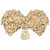
+ Open data
- Basic information
| Entry | Database: PDB / ID: 1eea | ||||||
|---|---|---|---|---|---|---|---|
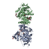
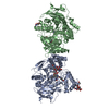
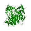
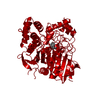
| Title | Acetylcholinesterase | ||||||
 Components Components | PROTEIN (ACETYLCHOLINESTERASE) | ||||||
 Keywords Keywords | HYDROLASE / SERINE HYDROLASE / ALPHA/BETA HYDROLASE / TETRAMER | ||||||
| Function / homology |  Function and homology information Function and homology informationacetylcholine catabolic process in synaptic cleft / choline metabolic process / acetylcholinesterase / acetylcholinesterase activity / synaptic cleft / side of membrane / synapse / extracellular space / plasma membrane Similarity search - Function | ||||||
| Biological species |  Electrophorus electricus (electric eel) Electrophorus electricus (electric eel) | ||||||
| Method |  X-RAY DIFFRACTION / X-RAY DIFFRACTION /  MOLECULAR REPLACEMENT / Resolution: 4.5 Å MOLECULAR REPLACEMENT / Resolution: 4.5 Å | ||||||
 Authors Authors | Raves, M.L. / Giles, K. / Schrag, J.D. / Schmid, M.F. / Phillips Jr., G.N. / Wah, C. / Howard, A.J. / Silman, I. / Sussman, J.L. | ||||||
 Citation Citation | Journal: Structure and Function of Cholinesterases and Related Proteins Year: 1998 Title: Quaternary Structure of Tetrameric Acetylcholinesterase Authors: Raves, M.L. / Giles, K. / Schrag, J.D. / Schmid, M.F. / Phillips Jr., G.N. / Wah, C. / Howard, A.J. / Silman, I. / Sussman, J.L. / Doctor, B.P. / Quinn, D.M. / Rotundo, R.L. / Taylor, P. #1: Journal: Protein Eng. / Year: 1997 Title: Interactions underlying subunit association in cholinesterases. Authors: Giles, K. #2: Journal: Science / Year: 1991 Title: Atomic structure of acetylcholinesterase from Torpedo californica: a prototypic acetylcholine-binding protein. Authors: Sussman, J.L. / Harel, M. / Frolow, F. / Oefner, C. / Goldman, A. / Toker, L. / Silman, I. #3: Journal: J.Biol.Chem. / Year: 1988 Title: Crystallization and preliminary X-ray diffraction analysis of 11 S acetylcholinesterase. Authors: Schrag, J.D. / Schmid, M.F. / Morgan, D.G. / Phillips Jr., G.N. / Chiu, W. / Tang, L. | ||||||
| History |
|
- Structure visualization
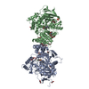
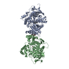
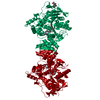
| Structure viewer | Molecule:  Molmil Molmil Jmol/JSmol Jmol/JSmol |
|---|
- Downloads & links
- Download
| PDBx/mmCIF format |  1eea.cif.gz 1eea.cif.gz | 102.7 KB | Display |  PDBx/mmCIF format PDBx/mmCIF format |
|---|---|---|---|---|
| PDB format |  pdb1eea.ent.gz pdb1eea.ent.gz | 76.1 KB | Display |  PDB format PDB format |
| PDBx/mmJSON format |  1eea.json.gz 1eea.json.gz | Tree view |  PDBx/mmJSON format PDBx/mmJSON format | |
| Others |  Other downloads Other downloads |
-Validation report
| Arichive directory |  https://data.pdbj.org/pub/pdb/validation_reports/ee/1eea https://data.pdbj.org/pub/pdb/validation_reports/ee/1eea ftp://data.pdbj.org/pub/pdb/validation_reports/ee/1eea ftp://data.pdbj.org/pub/pdb/validation_reports/ee/1eea | HTTPS FTP |
|---|
-Related structure data
| Related structure data | 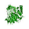 2aceS S: Starting model for refinement |
|---|---|
| Similar structure data |
- Links
Links
- Assembly
| Deposited unit | 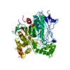
| ||||||||
|---|---|---|---|---|---|---|---|---|---|
| 1 | 
| ||||||||
| Unit cell |
|
- Components
| #1: Protein | Mass: 60461.195 Da / Num. of mol.: 1 / Source method: isolated from a natural source Details: SEE REMARK 450 FOR INFORMATION REGARDING THE SOURCE AND SEQUENCE Source: (natural)  Electrophorus electricus (electric eel) / Organ: ELECTRIC ORGAN / Variant: G4 FORM / Tissue: ELECTROPLAQUE / References: UniProt: P04058, acetylcholinesterase Electrophorus electricus (electric eel) / Organ: ELECTRIC ORGAN / Variant: G4 FORM / Tissue: ELECTROPLAQUE / References: UniProt: P04058, acetylcholinesterase |
|---|---|
| Has protein modification | Y |
| Source details | THE SEQUENCE LISTED IN THE SEQRES RECORD IS OF TORPEDO CALIFORNICA ACETYLCHOLINESTERASE, ...THE SEQUENCE LISTED IN THE SEQRES RECORD IS OF TORPEDO CALIFORNIC |
-Experimental details
-Experiment
| Experiment | Method:  X-RAY DIFFRACTION / Number of used crystals: 1 X-RAY DIFFRACTION / Number of used crystals: 1 |
|---|
- Sample preparation
| Crystal | Density Matthews: 6.5 Å3/Da / Density % sol: 81 % |
|---|---|
| Crystal grow | pH: 8 / Details: pH 8.0 |
-Data collection
| Diffraction | Mean temperature: 298 K |
|---|---|
| Diffraction source | Source:  ROTATING ANODE / Wavelength: 1.5418 ROTATING ANODE / Wavelength: 1.5418 |
| Detector | Type: XENTRONICS / Detector: AREA DETECTOR / Date: Jan 15, 1987 |
| Radiation | Protocol: SINGLE WAVELENGTH / Monochromatic (M) / Laue (L): M / Scattering type: x-ray |
| Radiation wavelength | Wavelength: 1.5418 Å / Relative weight: 1 |
| Reflection | Resolution: 4.5→34.3 Å / Num. obs: 9717 / % possible obs: 95.3 % / Observed criterion σ(I): 0 / Redundancy: 3.2 % / Rmerge(I) obs: 0.097 / Net I/σ(I): 6.7 |
| Reflection shell | Resolution: 4.5→4.65 Å / Redundancy: 1.7 % / Rmerge(I) obs: 0.272 / Mean I/σ(I) obs: 2 / % possible all: 75.2 |
- Processing
| Software |
| ||||||||||||
|---|---|---|---|---|---|---|---|---|---|---|---|---|---|
| Refinement | Method to determine structure:  MOLECULAR REPLACEMENT MOLECULAR REPLACEMENTStarting model: PDB ENTRY 2ACE Highest resolution: 4.5 Å Details: NO REFINEMENT WAS CARRIED OUT ON THE MODEL. FINAL MODEL IS MOLECULAR REPLACEMENT SOLUTION FOR SEARCH MODEL 2ACE. ONLY ONE MONOMER WAS FOUND TO OCCUPY THE ASYMMETRIC UNIT. THE FULL PACKING OF ...Details: NO REFINEMENT WAS CARRIED OUT ON THE MODEL. FINAL MODEL IS MOLECULAR REPLACEMENT SOLUTION FOR SEARCH MODEL 2ACE. ONLY ONE MONOMER WAS FOUND TO OCCUPY THE ASYMMETRIC UNIT. THE FULL PACKING OF THE UNIT CELL (16 MONOMERS, 4 TETRAMERS) SHOWS LARGE VOIDS THAT COULD EASILY BE OCCUPIED BY FOUR MORE TETRAMERS BY SIMPLE TRANSLATION OF THE OBTAINED TETRAMERS BY HALF THE C AXIS. REFINEMENT DOES NOT AGREE WITH TWO MONOMERS IN THE ASYMMETRIC UNIT. | ||||||||||||
| Refinement step | Cycle: LAST / Highest resolution: 4.5 Å
|
 Movie
Movie Controller












 PDBj
PDBj