 Movie
Movie Controller


+ Open data

- Basic information
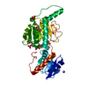
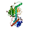
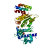
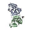
| Entry | Database: PDB / ID: 1e3o | ||||||
|---|---|---|---|---|---|---|---|
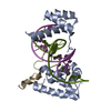
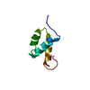
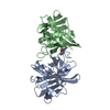
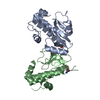
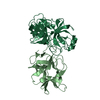
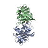
| Title | Crystal structure of Oct-1 POU dimer bound to MORE | ||||||
 Components Components |
| ||||||
 Keywords Keywords | TRANSCRIPTION / POU DOMAIN / DIMER / DNA BINDING | ||||||
| Function / homology |  Function and homology information Function and homology informationRNA Polymerase III Transcription Initiation From Type 3 Promoter / RNA Polymerase III Abortive And Retractive Initiation / RNA polymerase II transcribes snRNA genes / RNA polymerase II core promoter sequence-specific DNA binding / positive regulation of miRNA transcription / RNA polymerase II transcription regulator complex / Interleukin-4 and Interleukin-13 signaling / DNA-binding transcription activator activity, RNA polymerase II-specific / Estrogen-dependent gene expression / sequence-specific DNA binding ...RNA Polymerase III Transcription Initiation From Type 3 Promoter / RNA Polymerase III Abortive And Retractive Initiation / RNA polymerase II transcribes snRNA genes / RNA polymerase II core promoter sequence-specific DNA binding / positive regulation of miRNA transcription / RNA polymerase II transcription regulator complex / Interleukin-4 and Interleukin-13 signaling / DNA-binding transcription activator activity, RNA polymerase II-specific / Estrogen-dependent gene expression / sequence-specific DNA binding / DNA-binding transcription factor activity, RNA polymerase II-specific / RNA polymerase II cis-regulatory region sequence-specific DNA binding / negative regulation of DNA-templated transcription / regulation of transcription by RNA polymerase II / chromatin / endoplasmic reticulum / positive regulation of transcription by RNA polymerase II / DNA binding / nucleoplasm / identical protein binding / nucleus Similarity search - Function | ||||||
| Biological species |  Homo sapiens (human) Homo sapiens (human) | ||||||
| Method |  X-RAY DIFFRACTION / SYNCHROTRON / MOLECULAR REPLACEMENT / Resolution: 1.9 Å X-RAY DIFFRACTION / SYNCHROTRON / MOLECULAR REPLACEMENT / Resolution: 1.9 Å | ||||||
 Authors Authors | Remenyi, A. / Tomilin, A. / Pohl, E. / Schoeler, H. / Wilmanns, M. | ||||||
 Citation Citation | Journal: Mol.Cell / Year: 2001 Title: Differential Dimer Activities of the Transcription Factor Oct-1 by DNA-Induced Interface Swapping Authors: Remenyi, A. / Tomilin, A. / Pohl, E. / Lins, K. / Philippsen, A. / Reinbold, R. / Scholer, H.R. / Wilmanns, M. | ||||||
| History |
|
- Structure visualization
| Structure viewer | Molecule: MolmilJmol/JSmol |
|---|
- Downloads & links
-Download
| PDBx/mmCIF format | 1e3o.cif.gz | 57.7 KB | Display | PDBx/mmCIF format |
|---|---|---|---|---|
| PDB format | pdb1e3o.ent.gz | 38 KB | Display | PDB format |
| PDBx/mmJSON format | 1e3o.json.gz | Tree view | PDBx/mmJSON format | |
| Others |  Other downloads Other downloads |
-Validation report
| Arichive directory | https://data.pdbj.org/pub/pdb/validation_reports/e3/1e3oftp://data.pdbj.org/pub/pdb/validation_reports/e3/1e3o | HTTPS FTP |
|---|
-Related structure data
| Related structure data | 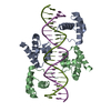 1hf0C 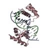 1octS S: Starting model for refinement C: citing same article ( |
|---|---|
| Similar structure data |


-Links
 PDBj
PDBj


- Assembly
| Deposited unit | 
| ||||||||
|---|---|---|---|---|---|---|---|---|---|
| 1 | 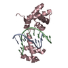
| ||||||||
| Unit cell |
| ||||||||
| Components on special symmetry positions |
|

-Components
| #1: DNA chain | Mass: 3422.260 Da / Num. of mol.: 1 / Source method: obtained synthetically / Source: (synth.) Homo sapiens (human) |
|---|---|
| #2: DNA chain | Mass: 3284.160 Da / Num. of mol.: 1 / Source method: obtained synthetically / Source: (synth.) Homo sapiens (human) |
| #3: Protein | Mass: 18398.877 Da / Num. of mol.: 1 / Fragment: DNA-BINDING DOMAIN / Mutation: YES Source method: isolated from a genetically manipulated source Source: (gene. exp.) Homo sapiens (human)Description: THE PROTEIN WAS EXPRESSED IN E.COLI BY RECOMBINANT DNA TECHNOLOGY Cellular location: NUCLEUS / Plasmid: PET-9D / Production host:  |
| #4: Water | ChemComp-HOH /  Mass: 18.015 Da / Num. of mol.: 138 / Source method: isolated from a natural source / Formula: H2O Mass: 18.015 Da / Num. of mol.: 138 / Source method: isolated from a natural source / Formula: H2O |
| Compound details | CHAIN C ENGINEERED |
-Experimental details
-Experiment
| Experiment | Method: X-RAY DIFFRACTION / Number of used crystals: 1 |
|---|
- Sample preparation
| Crystal | Density Matthews: 2.66 Å3/Da / Density % sol: 53.78 % | ||||||||||||||||||||
|---|---|---|---|---|---|---|---|---|---|---|---|---|---|---|---|---|---|---|---|---|---|
| Crystal grow | pH: 7 / Details: 22% PEG3350, 50MM HEPES, PH 7.0, 1.8MM SPERMINE | ||||||||||||||||||||
| Crystal grow | *PLUS Temperature: 20 ℃ / Method: vapor diffusion, hanging drop / Details: Remenyi, A., (2001) Acta Crystallogr., D57, 1634. | ||||||||||||||||||||
| Components of the solutions | *PLUS
|
-Data collection
| Diffraction | Mean temperature: 100 K |
|---|---|
| Diffraction source | Source: SYNCHROTRON / Site: EMBL/DESY, HAMBURG  / Beamline: BW7A / Wavelength: 0.8424 / Beamline: BW7A / Wavelength: 0.8424 |
| Detector | Type: MARRESEARCH / Detector: IMAGE PLATE / Date: Nov 15, 1999 |
| Radiation | Protocol: SINGLE WAVELENGTH / Monochromatic (M) / Laue (L): M / Scattering type: x-ray |
| Radiation wavelength | Wavelength: 0.8424 Å / Relative weight: 1 |
| Reflection | Resolution: 1.9→20 Å / Num. obs: 91810 / % possible obs: 95.3 % / Observed criterion σ(I): 2 / Redundancy: 4.6 % / Biso Wilson estimate: 36 Å2 / Rmerge(I) obs: 0.039 / Net I/σ(I): 24.4 |
| Reflection shell | Resolution: 1.9→1.97 Å / Rmerge(I) obs: 0.111 / Mean I/σ(I) obs: 6.2 / % possible all: 97 |
- Processing
| Software |
| ||||||||||||||||||||||||||||||||||||||||||||||||||||||||||||
|---|---|---|---|---|---|---|---|---|---|---|---|---|---|---|---|---|---|---|---|---|---|---|---|---|---|---|---|---|---|---|---|---|---|---|---|---|---|---|---|---|---|---|---|---|---|---|---|---|---|---|---|---|---|---|---|---|---|---|---|---|---|
| Refinement | Method to determine structure: MOLECULAR REPLACEMENT Starting model: 1OCT Resolution: 1.9→20 Å / Cross valid method: THROUGHOUT / σ(F): 0 Details: THE FOLLOWING AMINO ACID SIDE CHAINS A EXCLUDED FROM THE FINAL MODEL BECAUSE THEY HAVE POORLY DEFINED ELECTRON DENSITY: GLU 1, LYS 22, LYS 118, MET 121, LYS 125, GLU 129, LEU 133, GLU 136, LYS 142
| ||||||||||||||||||||||||||||||||||||||||||||||||||||||||||||
| Refinement step | Cycle: LAST / Resolution: 1.9→20 Å
| ||||||||||||||||||||||||||||||||||||||||||||||||||||||||||||
| Refine LS restraints |
| ||||||||||||||||||||||||||||||||||||||||||||||||||||||||||||
| Software | *PLUS Name: CNS / Version: 0.9 / Classification: refinement | ||||||||||||||||||||||||||||||||||||||||||||||||||||||||||||
| Refinement | *PLUS Lowest resolution: 20 Å / Rfactor obs: 0.22 / Rfactor Rwork: 0.22 | ||||||||||||||||||||||||||||||||||||||||||||||||||||||||||||
| Solvent computation | *PLUS | ||||||||||||||||||||||||||||||||||||||||||||||||||||||||||||
| Displacement parameters | *PLUS |
