 Movie
Movie Controller


+ Open data

- Basic information
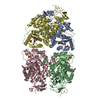
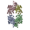
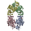
| Entry | Database: PDB / ID: 1e0u | ||||||
|---|---|---|---|---|---|---|---|
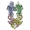
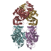
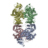
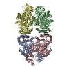
| Title | Structure R271L mutant of E. coli pyruvate kinase | ||||||
 Components Components | Pyruvate kinase | ||||||
 Keywords Keywords | PHOSPHOTRANSFERASE / GLYCOLYSIS / ALLOSTERY | ||||||
| Function / homology |  Function and homology information Function and homology informationpyruvate kinase complex / pyruvate kinase / pyruvate kinase activity / phosphorylation / potassium ion binding / glycolytic process / kinase activity / response to heat / magnesium ion binding / ATP binding ...pyruvate kinase complex / pyruvate kinase / pyruvate kinase activity / phosphorylation / potassium ion binding / glycolytic process / kinase activity / response to heat / magnesium ion binding / ATP binding / membrane / identical protein binding / cytosol / cytoplasm Similarity search - Function | ||||||
| Biological species |  | ||||||
| Method |  X-RAY DIFFRACTION / SYNCHROTRON / MOLECULAR REPLACEMENT / Resolution: 2.8 Å X-RAY DIFFRACTION / SYNCHROTRON / MOLECULAR REPLACEMENT / Resolution: 2.8 Å | ||||||
 Authors Authors | Fortin, R. / Mattevi, A. | ||||||
 Citation Citation | Journal: J.Biol.Chem. / Year: 2000 Title: The Allosteric Regulation of Pyruvate Kinase. Authors: Valentini, G. / Chiarelli, L. / Fortin, R. / Speranza, M.L. / Galizzi, A. / Mattevi, A. | ||||||
| History |
|
- Structure visualization
| Structure viewer | Molecule: MolmilJmol/JSmol |
|---|
- Downloads & links
-Download
| PDBx/mmCIF format | 1e0u.cif.gz | 355.8 KB | Display | PDBx/mmCIF format |
|---|---|---|---|---|
| PDB format | pdb1e0u.ent.gz | 283.6 KB | Display | PDB format |
| PDBx/mmJSON format | 1e0u.json.gz | Tree view | PDBx/mmJSON format | |
| Others |  Other downloads Other downloads |
-Validation report
| Arichive directory | https://data.pdbj.org/pub/pdb/validation_reports/e0/1e0uftp://data.pdbj.org/pub/pdb/validation_reports/e0/1e0u | HTTPS FTP |
|---|
-Related structure data
| Related structure data |  1e0tC  1pkyS S: Starting model for refinement C: citing same article ( |
|---|---|
| Similar structure data |

-Links
 PDBj
PDBj
- Assembly
| Deposited unit | 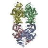
| ||||||||
|---|---|---|---|---|---|---|---|---|---|
| 1 |
| ||||||||
| Unit cell |
|
-Components
| #1: Protein | Mass: 50751.352 Da / Num. of mol.: 4 / Mutation: YES Source method: isolated from a genetically manipulated source Source: (gene. exp.) Gene: pykF, AC789_1c18560, ACN002_1349, EL75_1979, EL79_2019, EL80_2048, HMPREF3040_05259 Production host: References: UniProt: A0A0A0G552, UniProt: P0AD61*PLUS, pyruvate kinase #2: Chemical | ChemComp-SO4 /   Mass: 96.063 Da / Num. of mol.: 4 / Source method: obtained synthetically / Formula: SO4 Mass: 96.063 Da / Num. of mol.: 4 / Source method: obtained synthetically / Formula: SO4#3: Water | ChemComp-HOH / |  Mass: 18.015 Da / Num. of mol.: 200 / Source method: isolated from a natural source / Formula: H2O Mass: 18.015 Da / Num. of mol.: 200 / Source method: isolated from a natural source / Formula: H2OCompound details | CHAIN A, B, C, D ENGINEERED | |
|---|
-Experimental details
-Experiment
| Experiment | Method: X-RAY DIFFRACTION / Number of used crystals: 1 |
|---|
- Sample preparation
| Crystal | Density Matthews: 2.86 Å3/Da / Density % sol: 56.93 % | ||||||||||||||||||||||||||||||
|---|---|---|---|---|---|---|---|---|---|---|---|---|---|---|---|---|---|---|---|---|---|---|---|---|---|---|---|---|---|---|---|
| Crystal grow | pH: 6.2 / Details: pH 6.20 | ||||||||||||||||||||||||||||||
| Crystal grow | *PLUS pH: 6.5 / Method: vapor diffusion, hanging drop | ||||||||||||||||||||||||||||||
| Components of the solutions | *PLUS
|
-Data collection
| Diffraction | Mean temperature: 100 K |
|---|---|
| Diffraction source | Source: SYNCHROTRON / Site: ELETTRA  / Beamline: 5.2R / Wavelength: 1 / Beamline: 5.2R / Wavelength: 1 |
| Detector | Type: MARRESEARCH / Detector: IMAGE PLATE / Date: Feb 15, 1997 / Details: MIRRORS |
| Radiation | Protocol: SINGLE WAVELENGTH / Monochromatic (M) / Laue (L): M / Scattering type: x-ray |
| Radiation wavelength | Wavelength: 1 Å / Relative weight: 1 |
| Reflection | Resolution: 2.8→15 Å / Num. obs: 56604 / % possible obs: 97.7 % / Redundancy: 2.7 % / Rmerge(I) obs: 0.09 / Rsym value: 0.09 / Net I/σ(I): 6.5 |
| Reflection shell | Resolution: 2.8→2.9 Å / Redundancy: 2 % / Rmerge(I) obs: 0.201 / Mean I/σ(I) obs: 3.1 / Rsym value: 0.201 / % possible all: 86.1 |
| Reflection | *PLUS Num. measured all: 151954 |
| Reflection shell | *PLUS % possible obs: 86.1 % |
- Processing
| Software |
| |||||||||||||||||||||||||||||||||||||||||||||||||||||||||||||||
|---|---|---|---|---|---|---|---|---|---|---|---|---|---|---|---|---|---|---|---|---|---|---|---|---|---|---|---|---|---|---|---|---|---|---|---|---|---|---|---|---|---|---|---|---|---|---|---|---|---|---|---|---|---|---|---|---|---|---|---|---|---|---|---|---|
| Refinement | Method to determine structure: MOLECULAR REPLACEMENT Starting model: 1PKY Resolution: 2.8→15 Å / Cross valid method: THROUGHOUT / σ(F): 0
| |||||||||||||||||||||||||||||||||||||||||||||||||||||||||||||||
| Refinement step | Cycle: LAST / Resolution: 2.8→15 Å
| |||||||||||||||||||||||||||||||||||||||||||||||||||||||||||||||
| Refine LS restraints |
| |||||||||||||||||||||||||||||||||||||||||||||||||||||||||||||||
| Software | *PLUS Name: REFMAC / Classification: refinement | |||||||||||||||||||||||||||||||||||||||||||||||||||||||||||||||
| Refine LS restraints | *PLUS Type: p_plane_restr / Dev ideal: 0.009 |
