 Movie
Movie Controller

[English] 日本語
 Yorodumi
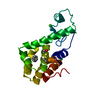
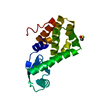
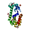
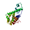
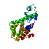
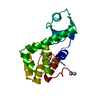
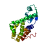
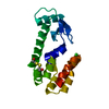
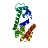
- PDB-1dyd: DETERMINATION OF ALPHA-HELIX PROPENSITY WITHIN THE CONTEXT OF A F... -
+ Open data

- Basic information
| Entry | Database: PDB / ID: 1dyd | ||||||
|---|---|---|---|---|---|---|---|
| Title | DETERMINATION OF ALPHA-HELIX PROPENSITY WITHIN THE CONTEXT OF A FOLDED PROTEIN: SITES 44 AND 131 IN BACTERIOPHAGE T4 LYSOZYME | ||||||
 Components Components | T4 LYSOZYME | ||||||
 Keywords Keywords | HYDROLASE(O-GLYCOSYL) | ||||||
| Function / homology |  Function and homology information Function and homology informationviral release from host cell by cytolysis / peptidoglycan catabolic process / cell wall macromolecule catabolic process / lysozyme / lysozyme activity / host cell cytoplasm / defense response to bacterium Similarity search - Function | ||||||
| Biological species |  Enterobacteria phage T4 (virus) Enterobacteria phage T4 (virus) | ||||||
| Method |  X-RAY DIFFRACTION / Resolution: 2.1 Å X-RAY DIFFRACTION / Resolution: 2.1 Å | ||||||
 Authors Authors | Zhang, X. / Matthews, B.W. | ||||||
 Citation Citation | Journal: J.Mol.Biol. / Year: 1994 Title: Determination of alpha-helix propensity within the context of a folded protein. Sites 44 and 131 in bacteriophage T4 lysozyme. Authors: Blaber, M. / Zhang, X.J. / Lindstrom, J.D. / Pepiot, S.D. / Baase, W.A. / Matthews, B.W. #1: Journal: To be PublishedTitle: Structural Basis of Amino Acid Alpha-Helix Propensity Authors: Blaber, M. / Zhang, X.-J. / Matthews, B.W. #2: Journal: Science / Year: 1989Title: Control of Enzyme Activity by an Engineered Disulfide Bond Authors: Matsumura, M. / Matthews, B.W. #3: Journal: J.Mol.Biol. / Year: 1987Title: Structure of Bacteriophage T4 Lysozyme Refined at 1.7 Angstroms Resolution Authors: Weaver, L.H. / Matthews, B.W. | ||||||
| History |
| ||||||
| Remark 700 | SHEET THERE ARE SEVERAL SUBTLE ASPECTS OF THE SECONDARY STRUCTURE OF THIS MOLECULE WHICH CANNOT ...SHEET THERE ARE SEVERAL SUBTLE ASPECTS OF THE SECONDARY STRUCTURE OF THIS MOLECULE WHICH CANNOT CONVENIENTLY BE REPRESENTED IN THE HELIX AND SHEET RECORDS BELOW. THESE ASPECTS INFLUENCE THE REPRESENTATION OF HELIX 6 AND STRAND 3 OF SHEET *S1*. THE PAPER J.MOL.BIOL., V. 118, P. 81, 1978 SHOULD BE CONSULTED FOR THESE SUBTLETIES. |
- Structure visualization
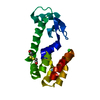
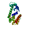
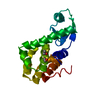
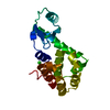
| Structure viewer | Molecule: MolmilJmol/JSmol |
|---|
- Downloads & links
-Download
| PDBx/mmCIF format | 1dyd.cif.gz | 48.6 KB | Display | PDBx/mmCIF format |
|---|---|---|---|---|
| PDB format | pdb1dyd.ent.gz | 34.1 KB | Display | PDB format |
| PDBx/mmJSON format | 1dyd.json.gz | Tree view | PDBx/mmJSON format | |
| Others |  Other downloads Other downloads |
-Validation report
| Arichive directory | https://data.pdbj.org/pub/pdb/validation_reports/dy/1dydftp://data.pdbj.org/pub/pdb/validation_reports/dy/1dyd | HTTPS FTP |
|---|
-Related structure data
| Related structure data |  1dyaC 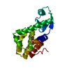 1dybC 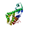 1dycC  1dyeC  1dyfC  1dygC 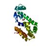 3l64C C: citing same article ( |
|---|---|
| Similar structure data |


-Links
 PDBj
PDBj


- Assembly
| Deposited unit | 
| ||||||||
|---|---|---|---|---|---|---|---|---|---|
| 1 |
| ||||||||
| Unit cell |
| ||||||||
| Atom site foot note | 1: SG SEO 97 IS BONDED TO SG CYS 97. |
-Components
| #1: Protein | Mass: 18676.494 Da / Num. of mol.: 1 Source method: isolated from a genetically manipulated source Source: (gene. exp.) Enterobacteria phage T4 (virus) / Genus: T4-like viruses / Species: Enterobacteria phage T4 sensu lato / Plasmid: M13 / References: UniProt: P00720, lysozyme | ||||||
|---|---|---|---|---|---|---|---|
| #2: Chemical |   Mass: 35.453 Da / Num. of mol.: 2 / Source method: obtained synthetically / Formula: Cl Mass: 35.453 Da / Num. of mol.: 2 / Source method: obtained synthetically / Formula: Cl#3: Chemical |   Mass: 78.133 Da / Num. of mol.: 2 / Source method: obtained synthetically / Formula: C2H6OS Mass: 78.133 Da / Num. of mol.: 2 / Source method: obtained synthetically / Formula: C2H6OS#4: Water | ChemComp-HOH / |  Mass: 18.015 Da / Num. of mol.: 145 / Source method: isolated from a natural source / Formula: H2O Mass: 18.015 Da / Num. of mol.: 145 / Source method: isolated from a natural source / Formula: H2OHas protein modification | N | |
-Experimental details
-Experiment
| Experiment | Method: X-RAY DIFFRACTION |
|---|
- Sample preparation
| Crystal | Density Matthews: 2.78 Å3/Da / Density % sol: 55.82 % | ||||||||||||||||||||||||||||||||||||||||||||||||||||||||
|---|---|---|---|---|---|---|---|---|---|---|---|---|---|---|---|---|---|---|---|---|---|---|---|---|---|---|---|---|---|---|---|---|---|---|---|---|---|---|---|---|---|---|---|---|---|---|---|---|---|---|---|---|---|---|---|---|---|
| Crystal grow | *PLUS Temperature: 4 ℃ / pH: 6.7 / Method: batch methodDetails: referred to 'Remington, S. J.', (1978) J. Mol. Biol., 118, 81-98 | ||||||||||||||||||||||||||||||||||||||||||||||||||||||||
| Components of the solutions | *PLUS
|
-Data collection
| Radiation | Scattering type: x-ray |
|---|---|
| Radiation wavelength | Relative weight: 1 |
| Reflection | *PLUS Highest resolution: 1.9 Å / Num. obs: 11796 / % possible obs: 74 % / Rmerge(I) obs: 0.088 |
- Processing
| Software | Name: TNT / Classification: refinement | ||||||||||||||||||||||||||||||
|---|---|---|---|---|---|---|---|---|---|---|---|---|---|---|---|---|---|---|---|---|---|---|---|---|---|---|---|---|---|---|---|
| Refinement | Resolution: 2.1→6 Å / σ(F): 0 Details: RESIDUES 162 - 164 IN WILD-TYPE AND ALL MUTANT LYSOZYMES ARE EXTREMELY MOBILE. THUS THE COORDINATES FOR THESE RESIDUES ARE VERY UNRELIABLE. THIS ENTRY DOES NOT INCLUDE RESIDUES 163 AND 164.
| ||||||||||||||||||||||||||||||
| Refinement step | Cycle: LAST / Resolution: 2.1→6 Å
| ||||||||||||||||||||||||||||||
| Refine LS restraints |
| ||||||||||||||||||||||||||||||
| Refinement | *PLUS Highest resolution: 2.1 Å / Lowest resolution: 6 Å / Num. reflection all: 11796 / σ(F): 0 / Rfactor all: 0.158 | ||||||||||||||||||||||||||||||
| Solvent computation | *PLUS | ||||||||||||||||||||||||||||||
| Displacement parameters | *PLUS | ||||||||||||||||||||||||||||||
| Refine LS restraints | *PLUS
|
