 Movie
Movie Controller

[English] 日本語
 Yorodumi
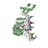
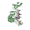
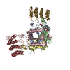
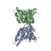
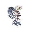
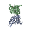
- PDB-1dto: CRYSTAL STRUCTURE OF THE COMPLETE TRANSACTIVATION DOMAIN OF E2 PR... -
+ Open data

- Basic information
| Entry | Database: PDB / ID: 1dto | ||||||
|---|---|---|---|---|---|---|---|
| Title | CRYSTAL STRUCTURE OF THE COMPLETE TRANSACTIVATION DOMAIN OF E2 PROTEIN FROM THE HUMAN PAPILLOMAVIRUS TYPE 16 | ||||||
 Components Components | REGULATORY PROTEIN E2 | ||||||
 Keywords Keywords | VIRAL PROTEIN / three-helix bundle / beta-sheet | ||||||
| Function / homology |  Function and homology information Function and homology informationhost cytoskeleton / viral DNA genome replication / regulation of DNA replication / DNA replication / DNA-binding transcription factor activity / nucleotide binding / DNA-templated transcription / host cell nucleus / DNA binding Similarity search - Function | ||||||
| Biological species |  Human papillomavirus type 16 Human papillomavirus type 16 | ||||||
| Method |  X-RAY DIFFRACTION / SYNCHROTRON / MIR / Resolution: 1.9 Å X-RAY DIFFRACTION / SYNCHROTRON / MIR / Resolution: 1.9 Å | ||||||
 Authors Authors | Antson, A.A. / Burns, J.E. / Moroz, O.V. / Scott, D.J. / Sanders, C.M. / Bronstein, I.B. / Dodson, G.G. / Wilson, K.S. / Maitland, N. | ||||||
 Citation Citation | Journal: Nature / Year: 2000 Title: Structure of the intact transactivation domain of the human papillomavirus E2 protein. Authors: Antson, A.A. / Burns, J.E. / Moroz, O.V. / Scott, D.J. / Sanders, C.M. / Bronstein, I.B. / Dodson, G.G. / Wilson, K.S. / Maitland, N.J. #1: Journal: Acta Crystallogr.,Sect.D / Year: 1998Title: Expression, Crystallisation and Preliminary X-ray Analysis of the E2 Transactivation Domain from Papillomavirus Type 16 Authors: Burns, J.E. / Moroz, O.V. / Antson, A.A. / Sanders, C.M. / Wilson, K.S. / Maitland, N.J. | ||||||
| History |
|
- Structure visualization
| Structure viewer | Molecule: MolmilJmol/JSmol |
|---|
- Downloads & links
-Download
| PDBx/mmCIF format | 1dto.cif.gz | 59.1 KB | Display | PDBx/mmCIF format |
|---|---|---|---|---|
| PDB format | pdb1dto.ent.gz | 42.6 KB | Display | PDB format |
| PDBx/mmJSON format | 1dto.json.gz | Tree view | PDBx/mmJSON format | |
| Others |  Other downloads Other downloads |
-Validation report
| Summary document | 1dto_validation.pdf.gz | 415 KB | Display | wwPDB validaton report |
|---|---|---|---|---|
| Full document | 1dto_full_validation.pdf.gz | 418.2 KB | Display | |
| Data in XML | 1dto_validation.xml.gz | 12.3 KB | Display | |
| Data in CIF | 1dto_validation.cif.gz | 17.8 KB | Display | |
| Arichive directory | https://data.pdbj.org/pub/pdb/validation_reports/dt/1dtoftp://data.pdbj.org/pub/pdb/validation_reports/dt/1dto | HTTPS FTP |
-Related structure data
| Similar structure data |
|---|


-Links
 PDBj
PDBj- Assembly
| Deposited unit | 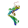
| ||||||||
|---|---|---|---|---|---|---|---|---|---|
| 1 | 
| ||||||||
| Unit cell |
| ||||||||
| Details | The biological assembly is a dimer constructed from chain A; the symmetry partner is generated by the crystallographic two-fold rotation X-Y,-Y,2/3-Z |
-Components
| #1: Protein | Mass: 25698.955 Da / Num. of mol.: 1 / Fragment: TRANSACTIVATION DOMAIN Source method: isolated from a genetically manipulated source Source: (gene. exp.) Human papillomavirus type 16 / Genus: Alphapapillomavirus / Species: Human papillomavirus - 16 / Plasmid: PET15B / Production host:  |
|---|---|
| #2: Water | ChemComp-HOH /  Mass: 18.015 Da / Num. of mol.: 211 / Source method: isolated from a natural source / Formula: H2O Mass: 18.015 Da / Num. of mol.: 211 / Source method: isolated from a natural source / Formula: H2O |
-Experimental details
-Experiment
| Experiment | Method: X-RAY DIFFRACTION / Number of used crystals: 1 |
|---|
- Sample preparation
| Crystal | Density Matthews: 2.4 Å3/Da / Density % sol: 55 % | ||||||||||||||||||||||||||||||||||||||||||||||||||||||||||||
|---|---|---|---|---|---|---|---|---|---|---|---|---|---|---|---|---|---|---|---|---|---|---|---|---|---|---|---|---|---|---|---|---|---|---|---|---|---|---|---|---|---|---|---|---|---|---|---|---|---|---|---|---|---|---|---|---|---|---|---|---|---|
| Crystal grow | Temperature: 291 K / Method: vapor diffusion, hanging drop / pH: 8.3 Details: monomethylether PEG 5000, NaCl, isopropanol, triethanolamine,Tris-HCl, DTT, EDTA, pH 8.3, VAPOR DIFFUSION, HANGING DROP, temperature 291.0K | ||||||||||||||||||||||||||||||||||||||||||||||||||||||||||||
| Crystal grow | *PLUS pH: 8 | ||||||||||||||||||||||||||||||||||||||||||||||||||||||||||||
| Components of the solutions | *PLUS
|
-Data collection
| Diffraction | Mean temperature: 120 K |
|---|---|
| Diffraction source | Source: SYNCHROTRON / Site: EMBL/DESY, HAMBURG  / Beamline: X11 / Wavelength: 0.89 / Beamline: X11 / Wavelength: 0.89 |
| Detector | Type: MARRESEARCH / Detector: IMAGE PLATE / Date: May 15, 1998 |
| Radiation | Protocol: SINGLE WAVELENGTH / Monochromatic (M) / Laue (L): M / Scattering type: x-ray |
| Radiation wavelength | Wavelength: 0.89 Å / Relative weight: 1 |
| Reflection | Resolution: 1.9→30 Å / Num. all: 21751 / Num. obs: 21751 / % possible obs: 98.8 % / Observed criterion σ(F): 0 / Observed criterion σ(I): 0 / Redundancy: 3.7 % / Biso Wilson estimate: 30.9 Å2 / Rmerge(I) obs: 0.058 / Net I/σ(I): 25.7 |
| Reflection shell | Resolution: 1.9→1.93 Å / Redundancy: 2.5 % / Rmerge(I) obs: 0.339 / Num. unique all: 837 / % possible all: 89.3 |
| Reflection | *PLUS |
| Reflection shell | *PLUS % possible obs: 89.3 % |
- Processing
| Software |
| ||||||||||||||||||||||||||||||||||||||||||||||||||||
|---|---|---|---|---|---|---|---|---|---|---|---|---|---|---|---|---|---|---|---|---|---|---|---|---|---|---|---|---|---|---|---|---|---|---|---|---|---|---|---|---|---|---|---|---|---|---|---|---|---|---|---|---|---|
| Refinement | Method to determine structure: MIR Starting model: NONE Resolution: 1.9→30 Å / SU B: 4.1 / SU ML: 0.121 / σ(F): 0 / σ(I): 0 / ESU R: 0.167 / ESU R Free: 0.176 / Stereochemistry target values: Engh & Huber
| ||||||||||||||||||||||||||||||||||||||||||||||||||||
| Refinement step | Cycle: LAST / Resolution: 1.9→30 Å
| ||||||||||||||||||||||||||||||||||||||||||||||||||||
| Refine LS restraints |
|
