 Movie
Movie Controller

[English] 日本語
 Yorodumi
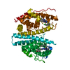
- PDB-1dqp: CRYSTAL STRUCTURE OF GIARDIA GUANINE PHOSPHORIBOSYLTRANSFERASE CO... -
+ Open data

- Basic information
| Entry | Database: PDB / ID: 1dqp | ||||||
|---|---|---|---|---|---|---|---|
| Title | CRYSTAL STRUCTURE OF GIARDIA GUANINE PHOSPHORIBOSYLTRANSFERASE COMPLEXED WITH IMMUCILLING | ||||||
 Components Components | GUANINE PHOSPHORIBOSYLTRANSFERASE | ||||||
 Keywords Keywords | TRANSFERASE / protein-inhibitor complex / immucillinG / 9-deazaguanine | ||||||
| Function / homology |  Function and homology information Function and homology informationguanine salvage / hypoxanthine metabolic process / hypoxanthine phosphoribosyltransferase activity / GMP salvage / IMP salvage / magnesium ion binding / cytosol Similarity search - Function | ||||||
| Biological species |  Giardia intestinalis (eukaryote) Giardia intestinalis (eukaryote) | ||||||
| Method |  X-RAY DIFFRACTION / SYNCHROTRON / Resolution: 1.75 Å X-RAY DIFFRACTION / SYNCHROTRON / Resolution: 1.75 Å | ||||||
 Authors Authors | Shi, W. / Munagala, N.R. / Wang, C.C. / Li, C.M. / Tyler, P.C. / Furneaux, R.H. / Grubmeyer, C. / Schramm, V.L. / Almo, S.C. | ||||||
 Citation Citation | Journal: Biochemistry / Year: 2000 Title: Crystal structures of Giardia lamblia guanine phosphoribosyltransferase at 1.75 A(,). Authors: Shi, W. / Munagala, N.R. / Wang, C.C. / Li, C.M. / Tyler, P.C. / Furneaux, R.H. / Grubmeyer, C. / Schramm, V.L. / Almo, S.C. | ||||||
| History |
|
- Structure visualization
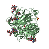
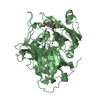
| Structure viewer | Molecule: MolmilJmol/JSmol |
|---|
- Downloads & links
-Download
| PDBx/mmCIF format | 1dqp.cif.gz | 113.9 KB | Display | PDBx/mmCIF format |
|---|---|---|---|---|
| PDB format | pdb1dqp.ent.gz | 88.4 KB | Display | PDB format |
| PDBx/mmJSON format | 1dqp.json.gz | Tree view | PDBx/mmJSON format | |
| Others |  Other downloads Other downloads |
-Validation report
| Arichive directory | https://data.pdbj.org/pub/pdb/validation_reports/dq/1dqpftp://data.pdbj.org/pub/pdb/validation_reports/dq/1dqp | HTTPS FTP |
|---|
-Related structure data


-Links
 PDBj
PDBj

- Assembly
| Deposited unit | 
| ||||||||
|---|---|---|---|---|---|---|---|---|---|
| 1 |
| ||||||||
| Unit cell |
| ||||||||
| Details | The biological assembly is a dimer in the asymmetric unit. |
-Components
| #1: Protein | Mass: 26367.279 Da / Num. of mol.: 2 Source method: isolated from a genetically manipulated source Source: (gene. exp.) Giardia intestinalis (eukaryote) / Production host:  References: UniProt: Q24973, hypoxanthine phosphoribosyltransferase #2: Chemical |   Mass: 281.268 Da / Num. of mol.: 2 / Source method: obtained synthetically / Formula: C11H15N5O4 Mass: 281.268 Da / Num. of mol.: 2 / Source method: obtained synthetically / Formula: C11H15N5O4#3: Chemical |   Mass: 60.095 Da / Num. of mol.: 2 / Source method: obtained synthetically / Formula: C3H8O Mass: 60.095 Da / Num. of mol.: 2 / Source method: obtained synthetically / Formula: C3H8O#4: Water | ChemComp-HOH / |  Mass: 18.015 Da / Num. of mol.: 485 / Source method: isolated from a natural source / Formula: H2O Mass: 18.015 Da / Num. of mol.: 485 / Source method: isolated from a natural source / Formula: H2ONonpolymer details | 9-deazaguanine is the only part of immucillinG ligand that could be seen in the density. Because ...9-deazaguanine is the only part of immucillinG ligand that could be seen in the density. Because immucillinG lacks a 5'-phosphate group, the sugar part of the ligand is not well ordered in this structure. | |
|---|
-Experimental details
-Experiment
| Experiment | Method: X-RAY DIFFRACTION / Number of used crystals: 1 |
|---|
- Sample preparation
| Crystal | Density Matthews: 2.36 Å3/Da / Density % sol: 47.92 % | |||||||||||||||||||||||||
|---|---|---|---|---|---|---|---|---|---|---|---|---|---|---|---|---|---|---|---|---|---|---|---|---|---|---|
| Crystal grow | Temperature: 296 K / Method: vapor diffusion, hanging drop / pH: 7.5 Details: 20% polyethylene glycol 4000, 5% isopropanol, 100 mM Hepes, pH 7.5, VAPOR DIFFUSION, HANGING DROP, temperature 296K | |||||||||||||||||||||||||
| Crystal grow | *PLUS Temperature: 18 ℃ | |||||||||||||||||||||||||
| Components of the solutions | *PLUS
|
-Data collection
| Diffraction | Mean temperature: 100 K |
|---|---|
| Diffraction source | Source: SYNCHROTRON / Site: NSLS  / Beamline: X9B / Wavelength: 0.979 / Beamline: X9B / Wavelength: 0.979 |
| Detector | Type: MARRESEARCH / Detector: CCD / Date: Aug 22, 1999 |
| Radiation | Protocol: SINGLE WAVELENGTH / Monochromatic (M) / Laue (L): M / Scattering type: x-ray |
| Radiation wavelength | Wavelength: 0.979 Å / Relative weight: 1 |
| Reflection | Resolution: 1.75→20 Å / Num. all: 50590 / Num. obs: 50590 / % possible obs: 98.9 % / Observed criterion σ(F): 0 / Observed criterion σ(I): 0 / Redundancy: 4.2 % / Biso Wilson estimate: 17 Å2 / Rsym value: 0.039 / Net I/σ(I): 40.8 |
| Reflection shell | Resolution: 1.75→1.78 Å / Redundancy: 3.6 % / Num. unique all: 2485 / Rsym value: 0.075 / % possible all: 98.7 |
| Reflection | *PLUS Num. measured all: 210690 / Rmerge(I) obs: 0.039 |
| Reflection shell | *PLUS % possible obs: 98.7 % / Rmerge(I) obs: 0.075 |
- Processing
| Software |
| |||||||||||||||||||||||||
|---|---|---|---|---|---|---|---|---|---|---|---|---|---|---|---|---|---|---|---|---|---|---|---|---|---|---|
| Refinement | Resolution: 1.75→20 Å / σ(F): 2 / σ(I): 1.4 / Stereochemistry target values: Engh & Huber / Details: Used weighted full matrix least squares procedure
| |||||||||||||||||||||||||
| Refinement step | Cycle: LAST / Resolution: 1.75→20 Å
| |||||||||||||||||||||||||
| Refine LS restraints |
| |||||||||||||||||||||||||
| Software | *PLUS Name: CNS / Version: 0.9 / Classification: refinement | |||||||||||||||||||||||||
| Refinement | *PLUS Num. reflection obs: 45122 | |||||||||||||||||||||||||
| Solvent computation | *PLUS | |||||||||||||||||||||||||
| Displacement parameters | *PLUS |
