 Movie
Movie Controller


+ Open data

- Basic information
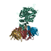
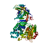
| Entry | Database: PDB / ID: 1dm0 | ||||||
|---|---|---|---|---|---|---|---|
| Title | SHIGA TOXIN | ||||||
 Components Components |
| ||||||
 Keywords Keywords | TOXIN / AB5 STRUCTURE / POLYPEPTIDE A / BLOCKING / ACTIVE SITE | ||||||
| Function / homology |  Function and homology information Function and homology informationhemolysis by symbiont of host erythrocytes / rRNA N-glycosylase / rRNA N-glycosylase activity / toxin activity / negative regulation of translation / extracellular region Similarity search - Function | ||||||
| Biological species |  Shigella dysenteriae (bacteria) Shigella dysenteriae (bacteria) | ||||||
| Method |  X-RAY DIFFRACTION / SYNCHROTRON / Resolution: 2.5 Å X-RAY DIFFRACTION / SYNCHROTRON / Resolution: 2.5 Å | ||||||
 Authors Authors | Fraser, M.E. / Chernaia, M.M. / Kozlov, Y.V. / James, M.N. | ||||||
 Citation Citation | Journal: Nat.Struct.Biol. / Year: 1994 Title: Crystal structure of the holotoxin from Shigella dysenteriae at 2.5 A resolution. Authors: Fraser, M.E. / Chernaia, M.M. / Kozlov, Y.V. / James, M.N. #1: Journal: Protein Toxin Structure, Parker, M.W., Ed. / Year: 1996Title: X-ray Crystal Structure of the Shiga Toxin Authors: Fraser, M.E. / Chernaia, M.M. / Kozlov, Y.V. / James, M.N. #2: Journal: J.Mol.Biol. / Year: 1993Title: Purification and Crystallization of Shiga Toxin from Shigella dysenteriae Authors: Kozlov, Y.V. / Chernaia, M.M. / Fraser, M.E. / James, M.N. | ||||||
| History |
|
- Structure visualization
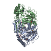
| Structure viewer | Molecule: MolmilJmol/JSmol |
|---|
- Downloads & links
-Download
| PDBx/mmCIF format | 1dm0.cif.gz | 234.4 KB | Display | PDBx/mmCIF format |
|---|---|---|---|---|
| PDB format | pdb1dm0.ent.gz | 197.8 KB | Display | PDB format |
| PDBx/mmJSON format | 1dm0.json.gz | Tree view | PDBx/mmJSON format | |
| Others |  Other downloads Other downloads |
-Validation report
| Summary document | 1dm0_validation.pdf.gz | 445.8 KB | Display | wwPDB validaton report |
|---|---|---|---|---|
| Full document | 1dm0_full_validation.pdf.gz | 508.5 KB | Display | |
| Data in XML | 1dm0_validation.xml.gz | 31.7 KB | Display | |
| Data in CIF | 1dm0_validation.cif.gz | 48.9 KB | Display | |
| Arichive directory | https://data.pdbj.org/pub/pdb/validation_reports/dm/1dm0ftp://data.pdbj.org/pub/pdb/validation_reports/dm/1dm0 | HTTPS FTP |
-Related structure data
| Similar structure data |
|---|


-Links
 PDBj
PDBj

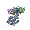
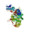
- Assembly
| Deposited unit | 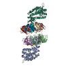
| ||||||||
|---|---|---|---|---|---|---|---|---|---|
| 1 | 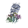
| ||||||||
| 2 | 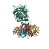
| ||||||||
| 3 |
| ||||||||
| Unit cell |
|
-Components
| #1: Protein | Mass: 31561.633 Da / Num. of mol.: 2 Source method: isolated from a genetically manipulated source Source: (gene. exp.) Shigella dysenteriae (bacteria) / Production host: References: UniProt: Q7BQ99, UniProt: Q9FBI2*PLUS, rRNA N-glycosylase #2: Protein | Mass: 7698.634 Da / Num. of mol.: 10 Source method: isolated from a genetically manipulated source Source: (gene. exp.) Shigella dysenteriae (bacteria) / Production host: #3: Water | ChemComp-HOH / |  Mass: 18.015 Da / Num. of mol.: 62 / Source method: isolated from a natural source / Formula: H2O Mass: 18.015 Da / Num. of mol.: 62 / Source method: isolated from a natural source / Formula: H2O |
|---|
-Experimental details
-Experiment
| Experiment | Method: X-RAY DIFFRACTION / Number of used crystals: 4 |
|---|
- Sample preparation
| Crystal | Density Matthews: 2.91 Å3/Da / Density % sol: 57.68 % |
|---|---|
| Crystal grow | Temperature: 294 K / Method: vapor diffusion, hanging drop / pH: 5 Details: sodium citrate, ethanol, pH 5, VAPOR DIFFUSION, HANGING DROP, temperature 294K |
| Crystal grow | *PLUS Details: Kozlov, Y.Z., (1993) J. Mol. Biol., 232, 704. |
-Data collection
| Diffraction | Mean temperature: 277 K |
|---|---|
| Diffraction source | Source: SYNCHROTRON / Site: Photon Factory  / Beamline: BL-6A / Wavelength: 1 / Beamline: BL-6A / Wavelength: 1 |
| Detector | Type: WEISSENBERG / Detector: DIFFRACTOMETER / Date: Oct 23, 1992 |
| Radiation | Protocol: SINGLE WAVELENGTH / Monochromatic (M) / Laue (L): M / Scattering type: x-ray |
| Radiation wavelength | Wavelength: 1 Å / Relative weight: 1 |
| Reflection | Resolution: 2.5→10 Å / Num. all: 47612 / Num. obs: 47612 / % possible obs: 83.1 % / Observed criterion σ(F): 0 / Observed criterion σ(I): 0 |
| Reflection shell | Resolution: 2.5→2.67 Å / Num. unique all: 5446 / % possible all: 53.1 |
| Reflection shell | *PLUS % possible obs: 53.1 % |
- Processing
| Software |
| ||||||||||||||||||||||||
|---|---|---|---|---|---|---|---|---|---|---|---|---|---|---|---|---|---|---|---|---|---|---|---|---|---|
| Refinement | Resolution: 2.5→10 Å / σ(F): 0 / σ(I): 0 / Stereochemistry target values: TNT dictionary / Details: Refinement with X-PLOR and TNT
| ||||||||||||||||||||||||
| Refinement step | Cycle: LAST / Resolution: 2.5→10 Å
| ||||||||||||||||||||||||
| Refine LS restraints |
| ||||||||||||||||||||||||
| Software | *PLUS Name: 'X-PLOR, TNT' / Classification: refinement | ||||||||||||||||||||||||
| Refine LS restraints | *PLUS
|
