 Movie
Movie Controller

[English] 日本語
 Yorodumi
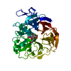
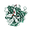
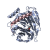
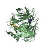
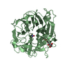
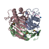
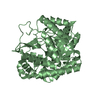
- PDB-1dim: SIALIDASE FROM SALMONELLA TYPHIMURIUM COMPLEXED WITH EPANA INHIBITOR -
+ Open data

- Basic information
| Entry | Database: PDB / ID: 1dim | ||||||
|---|---|---|---|---|---|---|---|
| Title | SIALIDASE FROM SALMONELLA TYPHIMURIUM COMPLEXED WITH EPANA INHIBITOR | ||||||
 Components Components | SIALIDASE | ||||||
 Keywords Keywords | GLYCOSIDASE / HYDROLASE | ||||||
| Function / homology |  Function and homology information Function and homology informationganglioside catabolic process / oligosaccharide catabolic process / exo-alpha-sialidase / exo-alpha-sialidase activity / membrane / cytoplasm Similarity search - Function | ||||||
| Biological species |  Salmonella typhimurium (bacteria) Salmonella typhimurium (bacteria) | ||||||
| Method |  X-RAY DIFFRACTION / DIFFERENCE MAP / Resolution: 1.6 Å X-RAY DIFFRACTION / DIFFERENCE MAP / Resolution: 1.6 Å | ||||||
 Authors Authors | Garman, E.F. / Crennell, S.C. / Vimr, E.R. / Laver, W.G. / Taylor, G.L. | ||||||
 Citation Citation | Journal: J.Mol.Biol. / Year: 1996 Title: The structures of Salmonella typhimurium LT2 neuraminidase and its complexes with three inhibitors at high resolution. Authors: Crennell, S.J. / Garman, E.F. / Philippon, C. / Vasella, A. / Laver, W.G. / Vimr, E.R. / Taylor, G.L. #1: Journal: Proc.Natl.Acad.Sci.USA / Year: 1993Title: Crystal Structure of a Bacterial Sialidase (from Salmonella Typhimurium Lt2) Shows the Same Fold as an Influenza Virus Neuraminidase Authors: Crennell, S.J. / Garman, E.F. / Laver, W.G. / Vimr, E.R. / Taylor, G.L. #2: Journal: J.Mol.Biol. / Year: 1992Title: Purification, Crystallization and Preliminary Crystallographic Study of Neuraminidase from Vibrio Cholerae and Salmonella Typhimurium Lt2 Authors: Taylor, G. / Vimr, E. / Garman, E. / Laver, G. #3: Journal: Helv.Chim.Acta / Year: 1990Title: Phosphonic-Acid Analogues of the N-Acetyl-2-Deoxyneuraminic Acids: Synthesis and Inhibition of Vibrio Cholerae Sialidase Authors: Wallimann, K. / Vasella, A. | ||||||
| History |
|
- Structure visualization
| Structure viewer | Molecule: MolmilJmol/JSmol |
|---|
- Downloads & links
-Download
| PDBx/mmCIF format | 1dim.cif.gz | 91 KB | Display | PDBx/mmCIF format |
|---|---|---|---|---|
| PDB format | pdb1dim.ent.gz | 68.1 KB | Display | PDB format |
| PDBx/mmJSON format | 1dim.json.gz | Tree view | PDBx/mmJSON format | |
| Others |  Other downloads Other downloads |
-Validation report
| Arichive directory | https://data.pdbj.org/pub/pdb/validation_reports/di/1dimftp://data.pdbj.org/pub/pdb/validation_reports/di/1dim | HTTPS FTP |
|---|
-Related structure data
| Related structure data |  1dilC  2silC  2simSC S: Starting model for refinement C: citing same article ( |
|---|---|
| Similar structure data |


-Links
 PDBj
PDBj

- Assembly
| Deposited unit | 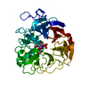
| ||||||||
|---|---|---|---|---|---|---|---|---|---|
| 1 |
| ||||||||
| Unit cell |
|
-Components
| #1: Protein | Mass: 41991.789 Da / Num. of mol.: 1 Source method: isolated from a genetically manipulated source Source: (gene. exp.) Salmonella typhimurium (bacteria) / Strain: LT2 / Description: RESIDUE MET 1 WAS EXCISED BY ESCHERICHIA COLI / Gene: NANH / Variant: TA263, PROTOTROPH / Plasmid: PSX62 / Gene (production host): NANH / Production host: |
|---|---|
| #2: Chemical | ChemComp-K /   Mass: 39.098 Da / Num. of mol.: 1 / Source method: obtained synthetically / Formula: K Mass: 39.098 Da / Num. of mol.: 1 / Source method: obtained synthetically / Formula: K |
| #3: Sugar | ChemComp-EQP / (  Type: D-saccharide / Mass: 329.241 Da / Num. of mol.: 1 / Source method: obtained synthetically / Formula: C10H20NO9P Type: D-saccharide / Mass: 329.241 Da / Num. of mol.: 1 / Source method: obtained synthetically / Formula: C10H20NO9P |
| #4: Water | ChemComp-HOH /  Mass: 18.015 Da / Num. of mol.: 219 / Source method: isolated from a natural source / Formula: H2O Mass: 18.015 Da / Num. of mol.: 219 / Source method: isolated from a natural source / Formula: H2O |
| Has protein modification | Y |
-Experimental details
-Experiment
| Experiment | Method: X-RAY DIFFRACTION / Number of used crystals: 1 |
|---|
- Sample preparation
| Crystal | Density Matthews: 2.13 Å3/Da / Density % sol: 42 % | ||||||||||||||||||||
|---|---|---|---|---|---|---|---|---|---|---|---|---|---|---|---|---|---|---|---|---|---|
| Crystal grow | *PLUS pH: 7.2 / Method: vapor diffusion, hanging drop | ||||||||||||||||||||
| Components of the solutions | *PLUS
|
-Data collection
| Diffraction | Mean temperature: 289 K |
|---|---|
| Diffraction source | Wavelength: 1.5418 |
| Detector | Type: SIEMENS / Detector: AREA DETECTOR / Date: Dec 5, 1992 |
| Radiation | Monochromator: GRAPHITE(002) / Monochromatic (M) / Laue (L): M / Scattering type: x-ray |
| Radiation wavelength | Wavelength: 1.5418 Å / Relative weight: 1 |
| Reflection | Resolution: 2.4→10 Å / Num. obs: 12091 / % possible obs: 89.6 % / Redundancy: 3.3 % / Rmerge(I) obs: 0.054 / Net I/σ(I): 14.8 |
| Reflection shell | Resolution: 1.6→1.66 Å / Redundancy: 2.7 % / Rmerge(I) obs: 0.078 / Mean I/σ(I) obs: 6.8 / % possible all: 87.8 |
| Reflection | *PLUS Lowest resolution: 9999 Å / Num. obs: 43572 / Num. measured all: 189373 |
| Reflection shell | *PLUS Lowest resolution: 1.7 Å |
- Processing
| Software |
| ||||||||||||||||||||||||||||||||||||||||||||||||||||||||||||||||||||||||||||||||
|---|---|---|---|---|---|---|---|---|---|---|---|---|---|---|---|---|---|---|---|---|---|---|---|---|---|---|---|---|---|---|---|---|---|---|---|---|---|---|---|---|---|---|---|---|---|---|---|---|---|---|---|---|---|---|---|---|---|---|---|---|---|---|---|---|---|---|---|---|---|---|---|---|---|---|---|---|---|---|---|---|---|
| Refinement | Method to determine structure: DIFFERENCE MAP Starting model: PDB ENTRY 2SIM Resolution: 1.6→6 Å / σ(F): 0 /
| ||||||||||||||||||||||||||||||||||||||||||||||||||||||||||||||||||||||||||||||||
| Displacement parameters | Biso mean: 14.42 Å2 | ||||||||||||||||||||||||||||||||||||||||||||||||||||||||||||||||||||||||||||||||
| Refinement step | Cycle: LAST / Resolution: 1.6→6 Å
| ||||||||||||||||||||||||||||||||||||||||||||||||||||||||||||||||||||||||||||||||
| Refine LS restraints |
| ||||||||||||||||||||||||||||||||||||||||||||||||||||||||||||||||||||||||||||||||
| Xplor file |
| ||||||||||||||||||||||||||||||||||||||||||||||||||||||||||||||||||||||||||||||||
| Software | *PLUS Name: X-PLOR / Classification: refinement | ||||||||||||||||||||||||||||||||||||||||||||||||||||||||||||||||||||||||||||||||
| Refinement | *PLUS | ||||||||||||||||||||||||||||||||||||||||||||||||||||||||||||||||||||||||||||||||
| Solvent computation | *PLUS | ||||||||||||||||||||||||||||||||||||||||||||||||||||||||||||||||||||||||||||||||
| Displacement parameters | *PLUS |
