 Movie
Movie Controller


+ Open data

- Basic information
| Entry | Database: PDB / ID: 1dbf | ||||||
|---|---|---|---|---|---|---|---|
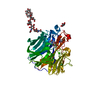
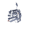
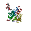
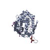
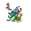
| Title | CHORISMATE MUTASE FROM BACILLUS SUBTILIS AT 1.30 ANGSTROM | ||||||
 Components Components | PROTEIN (CHORISMATE MUTASE) | ||||||
 Keywords Keywords | ISOMERASE / CHORISMATE MUTASE / SHIKIMATE PATHWAY | ||||||
| Function / homology |  Function and homology information Function and homology informationchorismate metabolic process / chorismate mutase / chorismate mutase activity / aromatic amino acid biosynthetic process / amino acid biosynthetic process / cytoplasm Similarity search - Function | ||||||
| Biological species |  | ||||||
| Method |  X-RAY DIFFRACTION / SYNCHROTRON / MOLECULAR REPLACEMENT / Resolution: 1.3 Å X-RAY DIFFRACTION / SYNCHROTRON / MOLECULAR REPLACEMENT / Resolution: 1.3 Å | ||||||
 Authors Authors | Gilliland, G.L. / Ladner, J.E. | ||||||
 Citation Citation | Journal: Acta Crystallogr.,Sect.D / Year: 2000 Title: The 1.30 A resolution structure of the Bacillus subtilis chorismate mutase catalytic homotrimer. Authors: Ladner, J.E. / Reddy, P. / Davis, A. / Tordova, M. / Howard, A.J. / Gilliland, G.L. | ||||||
| History |
|
- Structure visualization
| Structure viewer | Molecule: MolmilJmol/JSmol |
|---|
- Downloads & links
-Download
| PDBx/mmCIF format | 1dbf.cif.gz | 189.7 KB | Display | PDBx/mmCIF format |
|---|---|---|---|---|
| PDB format | pdb1dbf.ent.gz | 151.7 KB | Display | PDB format |
| PDBx/mmJSON format | 1dbf.json.gz | Tree view | PDBx/mmJSON format | |
| Others |  Other downloads Other downloads |
-Validation report
| Arichive directory | https://data.pdbj.org/pub/pdb/validation_reports/db/1dbfftp://data.pdbj.org/pub/pdb/validation_reports/db/1dbf | HTTPS FTP |
|---|
-Related structure data
| Related structure data | 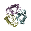 2chsS S: Starting model for refinement |
|---|---|
| Similar structure data |


-Links
 PDBj
PDBj
- Assembly
| Deposited unit | 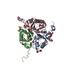
| ||||||||
|---|---|---|---|---|---|---|---|---|---|
| 1 |
| ||||||||
| Unit cell |
|
-Components
| #1: Protein | Mass: 14507.915 Da / Num. of mol.: 3 Source method: isolated from a genetically manipulated source Source: (gene. exp.) #2: Chemical | ChemComp-SO4 /   Mass: 96.063 Da / Num. of mol.: 9 / Source method: obtained synthetically / Formula: SO4 Mass: 96.063 Da / Num. of mol.: 9 / Source method: obtained synthetically / Formula: SO4#3: Chemical | ChemComp-GOL /   Mass: 92.094 Da / Num. of mol.: 5 / Source method: obtained synthetically / Formula: C3H8O3 Mass: 92.094 Da / Num. of mol.: 5 / Source method: obtained synthetically / Formula: C3H8O3#4: Water | ChemComp-HOH / |  Mass: 18.015 Da / Num. of mol.: 424 / Source method: isolated from a natural source / Formula: H2O Mass: 18.015 Da / Num. of mol.: 424 / Source method: isolated from a natural source / Formula: H2O |
|---|
-Experimental details
-Experiment
| Experiment | Method: X-RAY DIFFRACTION / Number of used crystals: 1 |
|---|
- Sample preparation
| Crystal | Density Matthews: 1.87 Å3/Da / Density % sol: 43.01 % Description: THE MODEL USED FOR MOLECULAR REPLACEMENT WAS ONE TRIMER (ABC) FROM STRUCTURE 2CHS. THE WATERS ARE GROUPED. WATERS 301-433 MAKE THEIR CLOSEST CONTACT WITH RESIDUES IN CHAIN A, WATERS 434- ...Description: THE MODEL USED FOR MOLECULAR REPLACEMENT WAS ONE TRIMER (ABC) FROM STRUCTURE 2CHS. THE WATERS ARE GROUPED. WATERS 301-433 MAKE THEIR CLOSEST CONTACT WITH RESIDUES IN CHAIN A, WATERS 434-567 WITH RESIDUES IN CHAIN B, WATERS 568-709 WITH RESIDUES IN CHAIN C, WATERS 710-717 WITH SO4 IONS AND WATERS 718- 724 WITH GLYCEROL MOLECULES. THE CLOSEST PROTEIN CHAIN FOR WATER MOLECULES 710, 711, 713, 715, 717, 718, 720, 722, 723, 724 IS CHAIN A. THE CLOSEST PROTEIN CHAIN FOR WATER MOLECULES 714 AND 719 IS CHAIN B. THE CLOSEST PROTEIN CHAIN FOR WATER MOLECULES 712, 716 AND 721 IS CHAIN C. | |||||||||||||||||||||||||||||||||||||||||||||
|---|---|---|---|---|---|---|---|---|---|---|---|---|---|---|---|---|---|---|---|---|---|---|---|---|---|---|---|---|---|---|---|---|---|---|---|---|---|---|---|---|---|---|---|---|---|---|
| Crystal grow | Temperature: 298 K / Method: vapor diffusion, hanging drop / pH: 3 Details: PROTEIN DROP: 5 MICROLITERS PROTEIN SOLUTION, 5 MICROLITERS RESERVOIR. PROTEIN SOLUTION: 13 MG/ML PROTEIN, 100 MM PMSF, 100 MM NACL, 50 MM TRIS PH 7.5, 1 MM EDTA, 1 MM DTT. RESERVOIR ...Details: PROTEIN DROP: 5 MICROLITERS PROTEIN SOLUTION, 5 MICROLITERS RESERVOIR. PROTEIN SOLUTION: 13 MG/ML PROTEIN, 100 MM PMSF, 100 MM NACL, 50 MM TRIS PH 7.5, 1 MM EDTA, 1 MM DTT. RESERVOIR SOLUTION: 2.2 M AMMONIUM SULFATE, 100 MM SODIUM ACETATE PH 3.0, VAPOR DIFFUSION, HANGING DROP, temperature 298K | |||||||||||||||||||||||||||||||||||||||||||||
| Crystal grow | *PLUS pH: 7.5 | |||||||||||||||||||||||||||||||||||||||||||||
| Components of the solutions | *PLUS
|
-Data collection
| Diffraction | Mean temperature: 100 K |
|---|---|
| Diffraction source | Source: SYNCHROTRON / Site: APS  / Beamline: 17-ID / Wavelength: 1 / Beamline: 17-ID / Wavelength: 1 |
| Detector | Type: BRUKER / Detector: CCD / Date: Feb 16, 1998 |
| Radiation | Protocol: SINGLE WAVELENGTH / Monochromatic (M) / Laue (L): M / Scattering type: x-ray |
| Radiation wavelength | Wavelength: 1 Å / Relative weight: 1 |
| Reflection | Num. obs: 89868 / % possible obs: 78 % / Redundancy: 2.1 % / Rmerge(I) obs: 0.092 / Net I/σ(I): 11.5 |
| Reflection shell | Resolution: 1.2→1.27 Å / Redundancy: 1.5 % / Rmerge(I) obs: 0.458 / Mean I/σ(I) obs: 1.2 / % possible all: 23 |
| Reflection | *PLUS Highest resolution: 1.3 Å / Lowest resolution: 100 Å / % possible obs: 92 % / Redundancy: 2.8 % / Num. measured all: 255732 |
| Reflection shell | *PLUS Highest resolution: 1.3 Å / Lowest resolution: 11.34 Å / % possible obs: 68 % / Redundancy: 1.7 % / Rmerge(I) obs: 0.336 |
- Processing
| Software |
| |||||||||||||||||||||||||||||||||
|---|---|---|---|---|---|---|---|---|---|---|---|---|---|---|---|---|---|---|---|---|---|---|---|---|---|---|---|---|---|---|---|---|---|---|
| Refinement | Method to determine structure: MOLECULAR REPLACEMENT Starting model: 2CHS Resolution: 1.3→100 Å / Num. parameters: 3296 / Num. restraintsaints: 4047 / Cross valid method: FREE R / σ(F): 0 / Stereochemistry target values: ENGH AND HUBER Details: THE EFFECTIVE RESOLUTION IS 1.30 ANGSTROMS, HOWEVER, ALL DATA AVAILABLE WERE USED DURING THE REFINEMENT. THIS INCLUDES SOME DATA AS HIGH AS 1.20 ANGSTROMS.
| |||||||||||||||||||||||||||||||||
| Solvent computation | Solvent model: MOEWS & KRETSINGER, J.MOL.BIOL.91(1973) 201-228 | |||||||||||||||||||||||||||||||||
| Refine analyze | Num. disordered residues: 3 / Occupancy sum hydrogen: 0 / Occupancy sum non hydrogen: 3529 | |||||||||||||||||||||||||||||||||
| Refinement step | Cycle: LAST / Resolution: 1.3→100 Å
| |||||||||||||||||||||||||||||||||
| Refine LS restraints |
|
