[English] 日本語
 Yorodumi
- PDB-1daq: SOLUTION STRUCTURE OF THE TYPE I DOCKERIN DOMAIN FROM THE CLOSTRI... -
+ Open data
- Basic information
| Entry | Database: PDB / ID: 1daq | ||||||
|---|---|---|---|---|---|---|---|
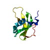
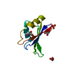
| Title | SOLUTION STRUCTURE OF THE TYPE I DOCKERIN DOMAIN FROM THE CLOSTRIDIUM THERMOCELLUM CELLULOSOME (MINIMIZED AVERAGE STRUCTURE) | ||||||
 Components Components | ENDOGLUCANASE SS | ||||||
 Keywords Keywords | HYDROLASE / CELLULOSE DEGRADATION / CELLULOSOME / CALCIUM-BINDING | ||||||
| Function / homology |  Function and homology information Function and homology informationcellulose 1,4-beta-cellobiosidase (reducing end) / cellulose 1,4-beta-cellobiosidase activity (reducing end) / cellulase activity / cellulose catabolic process / extracellular region / metal ion binding Similarity search - Function | ||||||
| Biological species |  Clostridium thermocellum (bacteria) Clostridium thermocellum (bacteria) | ||||||
| Method | SOLUTION NMR / torsion angle dynamics | ||||||
| Model type details | minimized average | ||||||
 Authors Authors | Lytle, B.L. / Volkman, B.F. / Westler, W.M. / Heckman, M.P. / Wu, J.H.D. | ||||||
 Citation Citation |  Journal: J.Mol.Biol. / Year: 2001 Journal: J.Mol.Biol. / Year: 2001Title: Solution structure of a type I dockerin domain, a novel prokaryotic, extracellular calcium-binding domain. Authors: Lytle, B.L. / Volkman, B.F. / Westler, W.M. / Heckman, M.P. / Wu, J.H. #1:  Journal: ARCH.BIOCHEM.BIOPHYS. / Year: 2000 Journal: ARCH.BIOCHEM.BIOPHYS. / Year: 2000Title: Secondary Structure and Calcium-induced Folding of the Clostridium thermocellum Dockerin Domain Determined by NMR Spectroscopy Authors: Lytle, B.L. / Volkman, B.F. / Westler, W.M. / Wu, J.H.D. #2:  Journal: J.Bacteriol. / Year: 1998 Journal: J.Bacteriol. / Year: 1998Title: Involvement of Both Dockerin Subdomains in Assembly of the Clostridium thermocellum Cellulosome Authors: Lytle, B. / Wu, J.H.D. | ||||||
| History |
| ||||||
| Remark 650 | HELIX DETERMINATION METHOD: AUTHOR-DETERMINED |
- Structure visualization
| Structure viewer | Molecule:  Molmil Molmil Jmol/JSmol Jmol/JSmol |
|---|
- Downloads & links
- Download
| PDBx/mmCIF format |  1daq.cif.gz 1daq.cif.gz | 33.4 KB | Display |  PDBx/mmCIF format PDBx/mmCIF format |
|---|---|---|---|---|
| PDB format |  pdb1daq.ent.gz pdb1daq.ent.gz | 22.5 KB | Display |  PDB format PDB format |
| PDBx/mmJSON format |  1daq.json.gz 1daq.json.gz | Tree view |  PDBx/mmJSON format PDBx/mmJSON format | |
| Others |  Other downloads Other downloads |
-Validation report
| Summary document |  1daq_validation.pdf.gz 1daq_validation.pdf.gz | 291.3 KB | Display |  wwPDB validaton report wwPDB validaton report |
|---|---|---|---|---|
| Full document |  1daq_full_validation.pdf.gz 1daq_full_validation.pdf.gz | 291.1 KB | Display | |
| Data in XML |  1daq_validation.xml.gz 1daq_validation.xml.gz | 3.5 KB | Display | |
| Data in CIF |  1daq_validation.cif.gz 1daq_validation.cif.gz | 4.2 KB | Display | |
| Arichive directory |  https://data.pdbj.org/pub/pdb/validation_reports/da/1daq https://data.pdbj.org/pub/pdb/validation_reports/da/1daq ftp://data.pdbj.org/pub/pdb/validation_reports/da/1daq ftp://data.pdbj.org/pub/pdb/validation_reports/da/1daq | HTTPS FTP |
-Related structure data
- Links
Links
- Assembly
| Deposited unit | 
| |||||||||
|---|---|---|---|---|---|---|---|---|---|---|
| 1 |
| |||||||||
| NMR ensembles |
|
- Components
| #1: Protein | Mass: 7858.770 Da / Num. of mol.: 1 / Fragment: TYPE I DOCKERIN DOMAIN (RESIDUES 673-741) Source method: isolated from a genetically manipulated source Source: (gene. exp.)  Clostridium thermocellum (bacteria) / Plasmid: PCYB2 / Production host: Clostridium thermocellum (bacteria) / Plasmid: PCYB2 / Production host:  References: UniProt: P38686, UniProt: P0C2S5*PLUS, cellulase |
|---|---|
| #2: Chemical |
-Experimental details
-Experiment
| Experiment | Method: SOLUTION NMR | ||||||||||||||||||||
|---|---|---|---|---|---|---|---|---|---|---|---|---|---|---|---|---|---|---|---|---|---|
| NMR experiment |
| ||||||||||||||||||||
| NMR details | Text: THIS STRUCTURE WAS DETERMINED BY STANDARD TECHNIQUES USING UNLABELED AND 15N- LABELED DOCKERIN. |
- Sample preparation
| Details | Contents: 100MM POTASSIUM CHLORIDE; 20MM CALCIUM CHLORIDE; 90% H2O, 10% D2O | |||||||||||||||
|---|---|---|---|---|---|---|---|---|---|---|---|---|---|---|---|---|
| Sample conditions |
| |||||||||||||||
| Crystal grow | *PLUS Method: other / Details: NMR |
-NMR measurement
| NMR spectrometer |
|
|---|
- Processing
| NMR software |
| ||||||||||||||||||||||||
|---|---|---|---|---|---|---|---|---|---|---|---|---|---|---|---|---|---|---|---|---|---|---|---|---|---|
| Refinement | Method: torsion angle dynamics / Software ordinal: 1 Details: THE STRUCTURE IS BASED ON 728 NOE-DERIVED DISTANCE CONSTRAINTS, 79 DIHEDRAL ANGLE CONSTRAINTS, AND 12 CALCIUM ION RESTRAINTS. | ||||||||||||||||||||||||
| NMR representative | Selection criteria: minimized average structure | ||||||||||||||||||||||||
| NMR ensemble | Conformers submitted total number: 1 |
 Movie
Movie Controller


 PDBj
PDBj


 NMRPipe