 Movie
Movie Controller


+ Open data

- Basic information
| Entry | Database: PDB / ID: 1cuk | ||||||
|---|---|---|---|---|---|---|---|
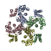
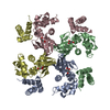
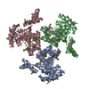
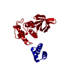
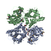
| Title | ESCHERICHIA COLI RUVA PROTEIN AT PH 4.9 AND ROOM TEMPERATURE | ||||||
 Components Components | RUVA PROTEIN | ||||||
 Keywords Keywords | HELICASE / DNA REPAIR / SOS RESPONSE / DNA-BINDING / DNA RECOMBINATION | ||||||
| Function / homology |  Function and homology information Function and homology informationHolliday junction helicase complex / Holliday junction resolvase complex / four-way junction helicase activity / SOS response / recombinational repair / four-way junction DNA binding / response to radiation / ATP binding / identical protein binding / cytoplasm Similarity search - Function | ||||||
| Biological species |  | ||||||
| Method |  X-RAY DIFFRACTION / Resolution: 1.9 Å X-RAY DIFFRACTION / Resolution: 1.9 Å | ||||||
 Authors Authors | Rafferty, J.B. / Rice, D.W. | ||||||
 Citation Citation | Journal: Science / Year: 1996 Title: Crystal structure of DNA recombination protein RuvA and a model for its binding to the Holliday junction. Authors: Rafferty, J.B. / Sedelnikova, S.E. / Hargreaves, D. / Artymiuk, P.J. / Baker, P.J. / Sharples, G.J. / Mahdi, A.A. / Lloyd, R.G. / Rice, D.W. #1: Journal: To be PublishedTitle: Crystallization of E.Coli Ruva Gives Insights Into the Symmetry of a Holliday Junction/Protein Complex Authors: Sedelnikova, S.E. / Rafferty, J.B. / Hargreaves, D. / Mahdi, A.A. / Lloyd, R.G. / Rice, D.W. | ||||||
| History |
|
- Structure visualization
| Structure viewer | Molecule: MolmilJmol/JSmol |
|---|
- Downloads & links
-Download
| PDBx/mmCIF format | 1cuk.cif.gz | 48.1 KB | Display | PDBx/mmCIF format |
|---|---|---|---|---|
| PDB format | pdb1cuk.ent.gz | 33.9 KB | Display | PDB format |
| PDBx/mmJSON format | 1cuk.json.gz | Tree view | PDBx/mmJSON format | |
| Others |  Other downloads Other downloads |
-Validation report
| Summary document | 1cuk_validation.pdf.gz | 368.4 KB | Display | wwPDB validaton report |
|---|---|---|---|---|
| Full document | 1cuk_full_validation.pdf.gz | 376.3 KB | Display | |
| Data in XML | 1cuk_validation.xml.gz | 6.5 KB | Display | |
| Data in CIF | 1cuk_validation.cif.gz | 9.3 KB | Display | |
| Arichive directory | https://data.pdbj.org/pub/pdb/validation_reports/cu/1cukftp://data.pdbj.org/pub/pdb/validation_reports/cu/1cuk | HTTPS FTP |
-Related structure data
| Similar structure data |
|---|

-Links
 PDBj
PDBj


- Assembly
| Deposited unit | 
| ||||||||
|---|---|---|---|---|---|---|---|---|---|
| 1 | 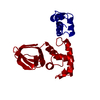
| ||||||||
| Unit cell |
|
-Components
| #1: Protein | Mass: 22197.783 Da / Num. of mol.: 1 Source method: isolated from a genetically manipulated source Source: (gene. exp.) |
|---|---|
| #2: Water | ChemComp-HOH /  Mass: 18.015 Da / Num. of mol.: 51 / Source method: isolated from a natural source / Formula: H2O Mass: 18.015 Da / Num. of mol.: 51 / Source method: isolated from a natural source / Formula: H2O |
-Experimental details
-Experiment
| Experiment | Method: X-RAY DIFFRACTION |
|---|
- Sample preparation
| Crystal | Density Matthews: 2.61 Å3/Da / Density % sol: 53 % | ||||||||||||||||||||
|---|---|---|---|---|---|---|---|---|---|---|---|---|---|---|---|---|---|---|---|---|---|
| Crystal grow | *PLUS Method: vapor diffusion, hanging dropDetails: Sedelnikova, S.E., (1999) Acta Crystallogr. D., 55, 263. PH range low: 7.5 / PH range high: 6.5 | ||||||||||||||||||||
| Components of the solutions | *PLUS
|
-Data collection
| Diffraction source | Wavelength: 1.5418 |
|---|---|
| Detector | Type: XUONG-HAMLIN MULTIWIRE / Detector: AREA DETECTOR / Date: Oct 26, 1995 |
| Radiation | Monochromatic (M) / Laue (L): M / Scattering type: x-ray |
| Radiation wavelength | Wavelength: 1.5418 Å / Relative weight: 1 |
| Reflection | Num. obs: 16391 / % possible obs: 89 % / Observed criterion σ(I): 0 / Redundancy: 2.7 % / Rmerge(I) obs: 0.073 |
- Processing
| Software |
| ||||||||||||||||||||||||||||||||||||||||
|---|---|---|---|---|---|---|---|---|---|---|---|---|---|---|---|---|---|---|---|---|---|---|---|---|---|---|---|---|---|---|---|---|---|---|---|---|---|---|---|---|---|
| Refinement | Resolution: 1.9→15 Å / σ(F): 0 Details: THE B-FACTORS FOR THE C-TERMINAL SEGMENT OF THE PROTEIN (RESIDUES 107 - 203) ARE HIGH WITH AN AVERAGE VALUE OF 73.6 A**2 OVER ALL ATOMS (71.0A**2 FOR MAIN CHAIN) AND THUS REPRESENT ...Details: THE B-FACTORS FOR THE C-TERMINAL SEGMENT OF THE PROTEIN (RESIDUES 107 - 203) ARE HIGH WITH AN AVERAGE VALUE OF 73.6 A**2 OVER ALL ATOMS (71.0A**2 FOR MAIN CHAIN) AND THUS REPRESENT POSITIONAL UNCERTAINTY OF APPROXIMATELY 1A IN SOME REGIONS.
| ||||||||||||||||||||||||||||||||||||||||
| Refinement step | Cycle: LAST / Resolution: 1.9→15 Å
| ||||||||||||||||||||||||||||||||||||||||
| Refine LS restraints |
| ||||||||||||||||||||||||||||||||||||||||
| Software | *PLUS Name: TNT / Classification: refinement | ||||||||||||||||||||||||||||||||||||||||
| Refinement | *PLUS Rfactor all: 0.209 / Rfactor Rfree: 0.027 | ||||||||||||||||||||||||||||||||||||||||
| Solvent computation | *PLUS | ||||||||||||||||||||||||||||||||||||||||
| Displacement parameters | *PLUS | ||||||||||||||||||||||||||||||||||||||||
| Refine LS restraints | *PLUS Type: t_plane_restr / Dev ideal: 0.008 / Weight: 5 |
