 Movie
Movie Controller

[English] 日本語
 Yorodumi
- PDB-1cty: MUTATION OF TYROSINE-67 IN CYTOCHROME C SIGNIFICANTLY ALTERS THE ... -
+ Open data

- Basic information
| Entry | Database: PDB / ID: 1cty | |||||||||
|---|---|---|---|---|---|---|---|---|---|---|
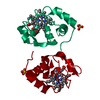
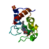
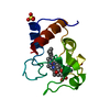
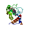
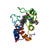
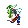
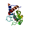
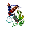
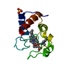
| Title | MUTATION OF TYROSINE-67 IN CYTOCHROME C SIGNIFICANTLY ALTERS THE LOCAL HEME ENVIRONMENT | |||||||||
 Components Components | CYTOCHROME C | |||||||||
 Keywords Keywords | ELECTRON TRANSPORT (HEME PROTEIN) | |||||||||
| Function / homology |  Function and homology information Function and homology informationRelease of apoptotic factors from the mitochondria / Pyroptosis / Detoxification of Reactive Oxygen Species / Respiratory electron transport / cardiolipin binding / mitochondrial electron transport, cytochrome c to oxygen / mitochondrial electron transport, ubiquinol to cytochrome c / mitochondrial intermembrane space / electron transfer activity / heme binding ...Release of apoptotic factors from the mitochondria / Pyroptosis / Detoxification of Reactive Oxygen Species / Respiratory electron transport / cardiolipin binding / mitochondrial electron transport, cytochrome c to oxygen / mitochondrial electron transport, ubiquinol to cytochrome c / mitochondrial intermembrane space / electron transfer activity / heme binding / mitochondrion / metal ion binding Similarity search - Function | |||||||||
| Biological species |  | |||||||||
| Method |  X-RAY DIFFRACTION / Resolution: 2.2 Å X-RAY DIFFRACTION / Resolution: 2.2 Å | |||||||||
 Authors Authors | Berghuis, A.M. / Brayer, G.D. | |||||||||
 Citation Citation | Journal: J.Mol.Biol. / Year: 1994 Title: Mutation of tyrosine-67 to phenylalanine in cytochrome c significantly alters the local heme environment. Authors: Berghuis, A.M. / Guillemette, J.G. / Smith, M. / Brayer, G.D. #1: Journal: J.Mol.Biol. / Year: 1992Title: Oxidation State-Dependent Conformational Changes in Cytochrome C Authors: Berghuis, A.M. / Brayer, G.D. #2: Journal: J.Mol.Biol. / Year: 1990Title: High-Resolution Refinement of Yeast Iso-1-Cytochrome C and Comparisons with Other Eukaryotic Cytochromes C Authors: Louie, G.V. / Brayer, G.D. #3: Journal: J.Mol.Biol. / Year: 1989Title: A Polypeptide Chain-Refolding Event Occurs in the Gly82 Variant of Yeast Iso-1-Cytochrome C Authors: Louie, G.V. / Brayer, G.D. #4: Journal: J.Mol.Biol. / Year: 1989Title: Crystallization of Yeast Iso-2-Cytochrome C Using a Novel Hair Seeding Technique Authors: Leung, C.J. / Nall, B.T. / Brayer, G.D. #5: Journal: J.Mol.Biol. / Year: 1988Title: Yeast Iso-1-Cytochrome C. A 2.8 Angstrom Resolution Three-Dimensional Structure Determination Authors: Louie, G.V. / Hutcheon, W.L.B. / Brayer, G.D. #6: Journal: Biochemistry / Year: 1988Title: Role of Phenylalanine-82 in Yeast Iso-1-Cytochrome C and Remote Conformational Changes Induced by a Serine Residue at This Position Authors: Louie, G.V. / Pielak, G.J. / Smith, M. / Brayer, G.D. | |||||||||
| History |
| |||||||||
| Remark 650 | HELIX THE END OF THE 50 HELIX (RESIDUE 55) IS DISTORTED. | |||||||||
| Remark 700 | SHEET RESIDUES IN SHEET S1 FORM A HIGHLY DISTORTED BETA TYPE CONFORMATION. |
- Structure visualization
| Structure viewer | Molecule: MolmilJmol/JSmol |
|---|
- Downloads & links
-Download
| PDBx/mmCIF format | 1cty.cif.gz | 38.5 KB | Display | PDBx/mmCIF format |
|---|---|---|---|---|
| PDB format | pdb1cty.ent.gz | 24.8 KB | Display | PDB format |
| PDBx/mmJSON format | 1cty.json.gz | Tree view | PDBx/mmJSON format | |
| Others |  Other downloads Other downloads |
-Validation report
| Arichive directory | https://data.pdbj.org/pub/pdb/validation_reports/ct/1ctyftp://data.pdbj.org/pub/pdb/validation_reports/ct/1cty | HTTPS FTP |
|---|
-Related structure data


-Links
 PDBj
PDBj


- Assembly
| Deposited unit | 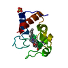
| ||||||||
|---|---|---|---|---|---|---|---|---|---|
| 1 |
| ||||||||
| Unit cell |
| ||||||||
| Atom site foot note | 1: RESIDUE LYS 72 AND TML 72 FORM EPSILON-N-TRIMETHYLLYSINE. SEE REMARK 9 ABOVE. 2: THE HEME GROUP IS COVALENTLY ATTACHED TO THE PROTEIN VIA THIOETHER BONDS TO THE SG ATOMS OF CYS 14 AND CYS 17. 3: IN ORDER TO CORRECTLY INDICATE IDENTICAL ATOMS BETWEEN THE COORDINATE ENTRIES OF THE MUTANT AND WILD-TYPE FORMS OF YEAST ISO-1 CYTOCHROME C (PROTEIN DATA BANK ENTRIES 1CTY AND 1YCC, RESPECTIVELY), ...3: IN ORDER TO CORRECTLY INDICATE IDENTICAL ATOMS BETWEEN THE COORDINATE ENTRIES OF THE MUTANT AND WILD-TYPE FORMS OF YEAST ISO-1 CYTOCHROME C (PROTEIN DATA BANK ENTRIES 1CTY AND 1YCC, RESPECTIVELY), THE SIDE-CHAIN ATOM NAMES OF SEVERAL RESIDUES CANNOT FOLLOW IUPAC/IUB SPECIFICATIONS WITH RESPECT TO THE DESIGNATION OF 1 AND 2 FOR BRANCHED SIDE CHAINS. THE AFFECTED RESIDUES ARE GLU -4, PHE -3, PHE 10, ARG 38, TYR 46, TYR 48, ASP 50, PHE 67, PHE 82, ASP 90, AND GLU 103. |
-Components
| #1: Protein | Mass: 12097.876 Da / Num. of mol.: 1 Source method: isolated from a genetically manipulated source Source: (gene. exp.) References: UniProt: P00044 |
|---|---|
| #2: Chemical | ChemComp-SO4 /   Mass: 96.063 Da / Num. of mol.: 1 / Source method: obtained synthetically / Formula: SO4 Mass: 96.063 Da / Num. of mol.: 1 / Source method: obtained synthetically / Formula: SO4 |
| #3: Chemical | ChemComp-HEC /   Mass: 618.503 Da / Num. of mol.: 1 / Source method: obtained synthetically / Formula: C34H34FeN4O4 Mass: 618.503 Da / Num. of mol.: 1 / Source method: obtained synthetically / Formula: C34H34FeN4O4 |
| #4: Water | ChemComp-HOH /  Mass: 18.015 Da / Num. of mol.: 30 / Source method: isolated from a natural source / Formula: H2O Mass: 18.015 Da / Num. of mol.: 30 / Source method: isolated from a natural source / Formula: H2O |
| Compound details | THIS PROTEIN HAS BEEN STABILIZED FOR ANALYSES BY THE MUTATION OF CYSTEINE 102 TO A THREONINE ...THIS PROTEIN HAS BEEN STABILIZED |
| Has protein modification | Y |
-Experimental details
-Experiment
| Experiment | Method: X-RAY DIFFRACTION |
|---|
- Sample preparation
| Crystal | Density Matthews: 1.92 Å3/Da / Density % sol: 35.85 % | ||||||||||||||||||||
|---|---|---|---|---|---|---|---|---|---|---|---|---|---|---|---|---|---|---|---|---|---|
| Crystal grow | *PLUS Method: vapor diffusion, hanging drop / Details: using a hair-seeding | ||||||||||||||||||||
| Components of the solutions | *PLUS
|
- Processing
| Software | Name: PROLSQ / Classification: refinement | ||||||||||||||||||||||||
|---|---|---|---|---|---|---|---|---|---|---|---|---|---|---|---|---|---|---|---|---|---|---|---|---|---|
| Refinement | Rfactor obs: 0.196 / Highest resolution: 2.2 Å | ||||||||||||||||||||||||
| Refinement step | Cycle: LAST / Highest resolution: 2.2 Å
| ||||||||||||||||||||||||
| Refine LS restraints |
| ||||||||||||||||||||||||
| Refinement | *PLUS Highest resolution: 2.2 Å / Rfactor obs: 0.196 | ||||||||||||||||||||||||
| Solvent computation | *PLUS | ||||||||||||||||||||||||
| Displacement parameters | *PLUS | ||||||||||||||||||||||||
| Refine LS restraints | *PLUS
|
