 Movie
Movie Controller

[English] 日本語
 Yorodumi
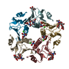
- PDB-1ct1: CHOLERA TOXIN B-PENTAMER MUTANT G33R BOUND TO RECEPTOR PENTASACCHARIDE -
+ Open data

- Basic information
| Entry | Database: PDB / ID: 1ct1 | |||||||||
|---|---|---|---|---|---|---|---|---|---|---|
| Title | CHOLERA TOXIN B-PENTAMER MUTANT G33R BOUND TO RECEPTOR PENTASACCHARIDE | |||||||||
 Components Components | CHOLERA TOXIN | |||||||||
 Keywords Keywords | ENTEROTOXIN / TOXIN-RECEPTOR COMPLEX / OLIGOSACCHARIDE | |||||||||
| Function / homology |  Function and homology information Function and homology informationgalactose binding / host cell surface binding / positive regulation of tyrosine phosphorylation of STAT protein / catalytic complex / toxin activity / periplasmic space / host cell plasma membrane / extracellular region Similarity search - Function | |||||||||
| Biological species |   Vibrio cholerae (bacteria) Vibrio cholerae (bacteria) | |||||||||
| Method |  X-RAY DIFFRACTION / Resolution: 2.3 Å X-RAY DIFFRACTION / Resolution: 2.3 Å | |||||||||
 Authors Authors | Merritt, E.A. / Hol, W.G.J. | |||||||||
 Citation Citation | Journal: Protein Sci. / Year: 1997 Title: Structural studies of receptor binding by cholera toxin mutants. Authors: Merritt, E.A. / Sarfaty, S. / Jobling, M.G. / Chang, T. / Holmes, R.K. / Hirst, T.R. / Hol, W.G. | |||||||||
| History |
|
- Structure visualization
| Structure viewer | Molecule: MolmilJmol/JSmol |
|---|
- Downloads & links
-Download
| PDBx/mmCIF format | 1ct1.cif.gz | 118.9 KB | Display | PDBx/mmCIF format |
|---|---|---|---|---|
| PDB format | pdb1ct1.ent.gz | 92.7 KB | Display | PDB format |
| PDBx/mmJSON format | 1ct1.json.gz | Tree view | PDBx/mmJSON format | |
| Others |  Other downloads Other downloads |
-Validation report
| Arichive directory | https://data.pdbj.org/pub/pdb/validation_reports/ct/1ct1ftp://data.pdbj.org/pub/pdb/validation_reports/ct/1ct1 | HTTPS FTP |
|---|
-Related structure data
| Related structure data |  2chbC  1chpS S: Starting model for refinement C: citing same article ( |
|---|---|
| Similar structure data |


-Links
 PDBj
PDBj
- Assembly
| Deposited unit | 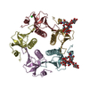
| ||||||||
|---|---|---|---|---|---|---|---|---|---|
| 1 |
| ||||||||
| Unit cell |
|
-Components
| #1: Protein | Mass: 11723.409 Da / Num. of mol.: 5 / Fragment: B-PENTAMER / Mutation: G33R Source method: isolated from a genetically manipulated source Source: (gene. exp.) Vibrio cholerae (bacteria) / Strain: OGAWA 41 (CLASSICAL BIOTYPE) / Production host: #2: Polysaccharide | Source method: isolated from a genetically manipulated source #3: Chemical | ChemComp-CL / |   Mass: 35.453 Da / Num. of mol.: 1 / Source method: obtained synthetically / Formula: Cl Mass: 35.453 Da / Num. of mol.: 1 / Source method: obtained synthetically / Formula: Cl#4: Water | ChemComp-HOH / |  Mass: 18.015 Da / Num. of mol.: 151 / Source method: isolated from a natural source / Formula: H2O Mass: 18.015 Da / Num. of mol.: 151 / Source method: isolated from a natural source / Formula: H2OCompound details | EACH CHAIN CONTAINS AN INTRODUCED | Has protein modification | Y | |
|---|
-Experimental details
-Experiment
| Experiment | Method: X-RAY DIFFRACTION / Number of used crystals: 1 |
|---|
- Sample preparation
| Crystal | Density Matthews: 2.26 Å3/Da / Density % sol: 45.69 % | ||||||||||||||||||||||||||||||||||||||||||||||||||||||||||||||||||||||||
|---|---|---|---|---|---|---|---|---|---|---|---|---|---|---|---|---|---|---|---|---|---|---|---|---|---|---|---|---|---|---|---|---|---|---|---|---|---|---|---|---|---|---|---|---|---|---|---|---|---|---|---|---|---|---|---|---|---|---|---|---|---|---|---|---|---|---|---|---|---|---|---|---|---|
| Crystal grow | Details: PROTEIN, 20 MM TRIS, 1 MM GM1-OS, PH 7.5 200 MM MGCL2, 100 MM CACODYLATE, 19% PEG 1000, 0.2% AGAROSE, PH 7.2 PH range: 7.2-7.5 | ||||||||||||||||||||||||||||||||||||||||||||||||||||||||||||||||||||||||
| Crystal grow | *PLUS pH: 7.5 / Method: vapor diffusion | ||||||||||||||||||||||||||||||||||||||||||||||||||||||||||||||||||||||||
| Components of the solutions | *PLUS
|
-Data collection
| Diffraction | Mean temperature: 287 K |
|---|---|
| Diffraction source | Source: ROTATING ANODE / Type: RIGAKU RUH2R / Wavelength: 1.5418 |
| Detector | Type: SIEMENS / Detector: AREA DETECTOR / Date: Dec 1, 1995 |
| Radiation | Monochromator: GRAPHITE(002) / Monochromatic (M) / Laue (L): M / Scattering type: x-ray |
| Radiation wavelength | Wavelength: 1.5418 Å / Relative weight: 1 |
| Reflection | Resolution: 2.3→15 Å / Num. obs: 22182 / % possible obs: 95 % / Rmerge(I) obs: 0.098 |
| Reflection shell | Resolution: 2.3→2.38 Å / % possible all: 55 |
| Reflection shell | *PLUS % possible obs: 55 % |
- Processing
| Software |
| ||||||||||||||||||||||||||||||||||||||||||||||||||||||||||||||||||||||||||||||||
|---|---|---|---|---|---|---|---|---|---|---|---|---|---|---|---|---|---|---|---|---|---|---|---|---|---|---|---|---|---|---|---|---|---|---|---|---|---|---|---|---|---|---|---|---|---|---|---|---|---|---|---|---|---|---|---|---|---|---|---|---|---|---|---|---|---|---|---|---|---|---|---|---|---|---|---|---|---|---|---|---|---|
| Refinement | Starting model: PDB ENTRY 1CHP Resolution: 2.3→15 Å / Data cutoff low absF: 0 / Isotropic thermal model: RESTRAINED / Cross valid method: THROUGHOUT / σ(F): 1 / Details: BABINET BULK SOLVENT MODEL KSOL = 0.8 BSOL = 50.0
| ||||||||||||||||||||||||||||||||||||||||||||||||||||||||||||||||||||||||||||||||
| Displacement parameters | Biso mean: 27 Å2
| ||||||||||||||||||||||||||||||||||||||||||||||||||||||||||||||||||||||||||||||||
| Refinement step | Cycle: LAST / Resolution: 2.3→15 Å
| ||||||||||||||||||||||||||||||||||||||||||||||||||||||||||||||||||||||||||||||||
| Refine LS restraints |
| ||||||||||||||||||||||||||||||||||||||||||||||||||||||||||||||||||||||||||||||||
| LS refinement shell | Resolution: 2.3→2.33 Å / Total num. of bins used: 15
| ||||||||||||||||||||||||||||||||||||||||||||||||||||||||||||||||||||||||||||||||
| Xplor file |
| ||||||||||||||||||||||||||||||||||||||||||||||||||||||||||||||||||||||||||||||||
| Software | *PLUS Name: X-PLOR / Version: 3.1 / Classification: refinement | ||||||||||||||||||||||||||||||||||||||||||||||||||||||||||||||||||||||||||||||||
| Refinement | *PLUS Rfactor Rfree: 0.25 | ||||||||||||||||||||||||||||||||||||||||||||||||||||||||||||||||||||||||||||||||
| Solvent computation | *PLUS | ||||||||||||||||||||||||||||||||||||||||||||||||||||||||||||||||||||||||||||||||
| Displacement parameters | *PLUS | ||||||||||||||||||||||||||||||||||||||||||||||||||||||||||||||||||||||||||||||||
| LS refinement shell | *PLUS Rfactor Rfree: 0.262 |
