 Movie
Movie Controller

[English] 日本語
 Yorodumi
- PDB-1csv: REPLACEMENTS IN A CONSERVED LEUCINE CLUSTER IN THE HYDROPHOBIC HE... -
+ Open data

- Basic information
| Entry | Database: PDB / ID: 1csv | |||||||||
|---|---|---|---|---|---|---|---|---|---|---|
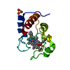
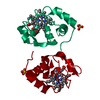
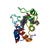
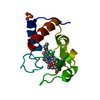
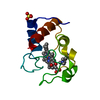
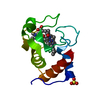
| Title | REPLACEMENTS IN A CONSERVED LEUCINE CLUSTER IN THE HYDROPHOBIC HEME POCKET OF CYTOCHROME C | |||||||||
 Components Components | CYTOCHROME C | |||||||||
 Keywords Keywords | ELECTRON TRANSPORT(HEME PROTEIN) | |||||||||
| Function / homology |  Function and homology information Function and homology informationRelease of apoptotic factors from the mitochondria / Pyroptosis / Detoxification of Reactive Oxygen Species / Respiratory electron transport / cardiolipin binding / mitochondrial electron transport, cytochrome c to oxygen / mitochondrial electron transport, ubiquinol to cytochrome c / mitochondrial intermembrane space / electron transfer activity / heme binding ...Release of apoptotic factors from the mitochondria / Pyroptosis / Detoxification of Reactive Oxygen Species / Respiratory electron transport / cardiolipin binding / mitochondrial electron transport, cytochrome c to oxygen / mitochondrial electron transport, ubiquinol to cytochrome c / mitochondrial intermembrane space / electron transfer activity / heme binding / mitochondrion / metal ion binding Similarity search - Function | |||||||||
| Biological species |  | |||||||||
| Method |  X-RAY DIFFRACTION / Resolution: 1.9 Å X-RAY DIFFRACTION / Resolution: 1.9 Å | |||||||||
 Authors Authors | Lo, T.P. / Brayer, G.D. | |||||||||
 Citation Citation | Journal: Protein Sci. / Year: 1995 Title: Replacements in a conserved leucine cluster in the hydrophobic heme pocket of cytochrome c. Authors: Lo, T.P. / Murphy, M.E. / Guillemette, J.G. / Smith, M. / Brayer, G.D. #1: Journal: Biochemistry / Year: 1995Title: Structural Studies of the Roles of Residues 82 and 85 at the Interactive Face of Cytochrome C Authors: Lo, T.P. / Guillemette, J.G. / Louie, G.V. / Smith, M. / Brayer, G.D. #2: Journal: J.Mol.Biol. / Year: 1992Title: Oxidation State-Dependent Conformational Changes in Cytochrome C Authors: Berghuis, A.M. / Brayer, G.D. #3: Journal: J.Mol.Biol. / Year: 1990Title: High-Resolution Refinement of Yeast Iso-1-Cytochrome C and Comparisons with Other Eukaryotic Cytochromes C Authors: Louie, G.V. / Brayer, G.D. #4: Journal: J.Mol.Biol. / Year: 1989Title: A Polypeptide Chain-Refolding Event Occurs in the Gly82 Variant of Yeast Iso-1-Cytochrome C Authors: Louie, G.V. / Brayer, G.D. #5: Journal: J.Mol.Biol. / Year: 1989Title: Crystallization of Yeast Iso-2-Cytochrome C Using a Novel Hair Seeding Technique Authors: Leung, C.J. / Nall, B.T. / Brayer, G.D. #6: Journal: J.Mol.Biol. / Year: 1988Title: Yeast Iso-1-Cytochrome C. A 2.8 Angstrom Resolution Three-Dimensional Structure Determination Authors: Louie, G.V. / Hutcheon, W.L.B. / Brayer, G.D. #7: Journal: Biochemistry / Year: 1988Title: Role of Phenylalanine-82 in Yeast Iso-1-Cytochrome C and Remote Conformational Changes Induced by a Serine Residue at This Position Authors: Louie, G.V. / Pielak, G.J. / Smith, M. / Brayer, G.D. | |||||||||
| History |
|
- Structure visualization
| Structure viewer | Molecule: MolmilJmol/JSmol |
|---|
- Downloads & links
-Download
| PDBx/mmCIF format | 1csv.cif.gz | 38.9 KB | Display | PDBx/mmCIF format |
|---|---|---|---|---|
| PDB format | pdb1csv.ent.gz | 25 KB | Display | PDB format |
| PDBx/mmJSON format | 1csv.json.gz | Tree view | PDBx/mmJSON format | |
| Others |  Other downloads Other downloads |
-Validation report
| Arichive directory | https://data.pdbj.org/pub/pdb/validation_reports/cs/1csvftp://data.pdbj.org/pub/pdb/validation_reports/cs/1csv | HTTPS FTP |
|---|
-Related structure data


-Links
 PDBj
PDBj


- Assembly
| Deposited unit | 
| ||||||||
|---|---|---|---|---|---|---|---|---|---|
| 1 |
| ||||||||
| Unit cell |
| ||||||||
| Atom site foot note | 1: THE HEME GROUP IS COVALENTLY ATTACHED TO THE PROTEIN VIA THIOETHER BONDS FROM THE SG ATOMS OF CYS 14 AND CYS 17, TO THE CAB AND CAC HEME ATOMS, RESPECTIVELY. 2: RESIDUE 72 IS EPSILON-N-TRIMETHYLLYSINE. THE THREE METHYL CARBONS FOR THIS RESIDUE ARE PRESENT AS HETATMS WITH RESIDUE NAME TML (FOLLOWING THE HEME). RESIDUE 72 IS IDENTIFIED AS LYS ON THE ATOM ...2: RESIDUE 72 IS EPSILON-N-TRIMETHYLLYSINE. THE THREE METHYL CARBONS FOR THIS RESIDUE ARE PRESENT AS HETATMS WITH RESIDUE NAME TML (FOLLOWING THE HEME). RESIDUE 72 IS IDENTIFIED AS LYS ON THE ATOM AND SEQRES RECORDS. RESIDUE LYS 72 IS TRIMETHYLATED AT THE AMINO END OF ITS SIDE CHAIN. 3: RESIDUES MET 80 AND HIS 18 FORM HEME IRON LIGAND BONDS. |
-Components
| #1: Protein | Mass: 12147.893 Da / Num. of mol.: 1 Source method: isolated from a genetically manipulated source Source: (gene. exp.) References: UniProt: P00044 | ||
|---|---|---|---|
| #2: Chemical | ChemComp-SO4 /   Mass: 96.063 Da / Num. of mol.: 1 / Source method: obtained synthetically / Formula: SO4 Mass: 96.063 Da / Num. of mol.: 1 / Source method: obtained synthetically / Formula: SO4 | ||
| #3: Chemical | ChemComp-HEC /   Mass: 618.503 Da / Num. of mol.: 1 / Source method: obtained synthetically / Formula: C34H34FeN4O4 Mass: 618.503 Da / Num. of mol.: 1 / Source method: obtained synthetically / Formula: C34H34FeN4O4 | ||
| #4: Water | ChemComp-HOH /  Mass: 18.015 Da / Num. of mol.: 58 / Source method: isolated from a natural source / Formula: H2O Mass: 18.015 Da / Num. of mol.: 58 / Source method: isolated from a natural source / Formula: H2O | ||
| Compound details | THIS PROTEIN HAS BEEN STABILIZED| Has protein modification | Y | |
-Experimental details
-Experiment
| Experiment | Method: X-RAY DIFFRACTION |
|---|
- Sample preparation
| Crystal | Density Matthews: 1.91 Å3/Da / Density % sol: 35.47 % | |||||||||||||||||||||||||
|---|---|---|---|---|---|---|---|---|---|---|---|---|---|---|---|---|---|---|---|---|---|---|---|---|---|---|
| Crystal grow | *PLUS pH: 6.7 / Method: vapor diffusion, hanging drop | |||||||||||||||||||||||||
| Components of the solutions | *PLUS
|
-Data collection
| Radiation | Scattering type: x-ray |
|---|---|
| Radiation wavelength | Relative weight: 1 |
| Reflection | *PLUS Highest resolution: 1.9 Å / Num. measured all: 8072 |
- Processing
| Software | Name: PROLSQ / Classification: refinement | ||||||||||||||||||||||||||||||||||||||||||||||||||||||||||||||||||||||||||||||||||||
|---|---|---|---|---|---|---|---|---|---|---|---|---|---|---|---|---|---|---|---|---|---|---|---|---|---|---|---|---|---|---|---|---|---|---|---|---|---|---|---|---|---|---|---|---|---|---|---|---|---|---|---|---|---|---|---|---|---|---|---|---|---|---|---|---|---|---|---|---|---|---|---|---|---|---|---|---|---|---|---|---|---|---|---|---|---|
| Refinement | Resolution: 1.9→6 Å / σ(F): 2 /
| ||||||||||||||||||||||||||||||||||||||||||||||||||||||||||||||||||||||||||||||||||||
| Displacement parameters | Biso mean: 16.5 Å2 | ||||||||||||||||||||||||||||||||||||||||||||||||||||||||||||||||||||||||||||||||||||
| Refinement step | Cycle: LAST / Resolution: 1.9→6 Å
| ||||||||||||||||||||||||||||||||||||||||||||||||||||||||||||||||||||||||||||||||||||
| Refine LS restraints |
|
