 Movie
Movie Controller


+ Open data

- Basic information
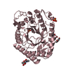
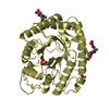
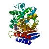
| Entry | Database: PDB / ID: 1cef | |||||||||
|---|---|---|---|---|---|---|---|---|---|---|
| Title | CEFOTAXIME COMPLEXED WITH THE STREPTOMYCES R61 DD-PEPTIDASE | |||||||||
 Components Components | D-ALANYL-D-ALANINE CARBOXYPEPTIDASE TRANSPEPTIDASE | |||||||||
 Keywords Keywords | HYDROLASE-TRANSPEPTIDASE / D-AMINO ACID PEPTIDASE / PENICILLIN TARGET | |||||||||
| Function / homology |  Function and homology information Function and homology informationserine-type D-Ala-D-Ala carboxypeptidase / serine-type D-Ala-D-Ala carboxypeptidase activity / peptidoglycan biosynthetic process / cell wall organization / regulation of cell shape / proteolysis / extracellular region Similarity search - Function | |||||||||
| Biological species |  Streptomyces sp. (bacteria) Streptomyces sp. (bacteria) | |||||||||
| Method |  X-RAY DIFFRACTION / Resolution: 2.04 Å X-RAY DIFFRACTION / Resolution: 2.04 Å | |||||||||
 Authors Authors | Knox, J.R. / Kuzin, A.P. | |||||||||
 Citation Citation | Journal: Biochemistry / Year: 1995 Title: Binding of cephalothin and cefotaxime to D-ala-D-ala-peptidase reveals a functional basis of a natural mutation in a low-affinity penicillin-binding protein and in extended-spectrum beta-lactamases. Authors: Kuzin, A.P. / Liu, H. / Kelly, J.A. / Knox, J.R. #1: Journal: J.Mol.Biol. / Year: 1995Title: The Refined Crystallographic Structure of a Dd-Peptidase Penicillin-Target Enzyme at 1.6 A Resolution Authors: Kelly, J.A. / Kuzin, A.P. #2: Journal: J.Mol.Biol. / Year: 1989Title: Crystallographic Mapping of Beta-Lactams Bound to a D-Alanyl-D-Alanine Peptidase Target Enzyme Authors: Kelly, J.A. / Knox, J.R. / Zhao, H. / Frere, J.M. / Ghaysen, J.M. #3: Journal: Science / Year: 1986 Title: On the origin of bacterial resistance to penicillin: comparison of a beta-lactamase and a penicillin target. Authors: Kelly, J.A. / Dideberg, O. / Charlier, P. / Wery, J.P. / Libert, M. / Moews, P.C. / Knox, J.R. / Duez, C. / Fraipont, C. / Joris, B. #4: Journal: J.Biol.Chem. / Year: 1985Title: 2.8-A Structure of Penicillin-Sensitive D-Alanyl Carboxypeptidase-Transpeptidase from Streptomyces R61 and Complexes with Beta-Lactams Authors: Kelly, J.A. / Knox, J.R. / Moews, P.C. / Hite, G.J. / Bartolone, J.B. / Zhao, H. / Joris, B. / Frere, J.M. / Ghuysen, J.M. | |||||||||
| History |
|
- Structure visualization
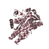
| Structure viewer | Molecule: MolmilJmol/JSmol |
|---|
- Downloads & links
-Download
| PDBx/mmCIF format | 1cef.cif.gz | 82.1 KB | Display | PDBx/mmCIF format |
|---|---|---|---|---|
| PDB format | pdb1cef.ent.gz | 60.7 KB | Display | PDB format |
| PDBx/mmJSON format | 1cef.json.gz | Tree view | PDBx/mmJSON format | |
| Others |  Other downloads Other downloads |
-Validation report
| Arichive directory | https://data.pdbj.org/pub/pdb/validation_reports/ce/1cefftp://data.pdbj.org/pub/pdb/validation_reports/ce/1cef | HTTPS FTP |
|---|
-Related structure data


-Links
 PDBj
PDBj
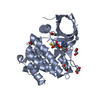
- Assembly
| Deposited unit | 
| ||||||||
|---|---|---|---|---|---|---|---|---|---|
| 1 |
| ||||||||
| Unit cell |
|
-Components
| #1: Protein | Mass: 37422.574 Da / Num. of mol.: 1 / Source method: isolated from a natural source / Source: (natural) Streptomyces sp. (bacteria) / Strain: R61References: UniProt: P15555, serine-type D-Ala-D-Ala carboxypeptidase |
|---|---|
| #2: Chemical | ChemComp-CEF / 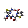  Mass: 397.429 Da / Num. of mol.: 1 / Source method: obtained synthetically / Formula: C14H15N5O5S2 Mass: 397.429 Da / Num. of mol.: 1 / Source method: obtained synthetically / Formula: C14H15N5O5S2 |
| #3: Water | ChemComp-HOH /  Mass: 18.015 Da / Num. of mol.: 246 / Source method: isolated from a natural source / Formula: H2O Mass: 18.015 Da / Num. of mol.: 246 / Source method: isolated from a natural source / Formula: H2O |
| Has protein modification | Y |
-Experimental details
-Experiment
| Experiment | Method: X-RAY DIFFRACTION |
|---|
- Sample preparation
| Crystal | Density Matthews: 2.37 Å3/Da / Density % sol: 48.11 % | |||||||||||||||||||||||||
|---|---|---|---|---|---|---|---|---|---|---|---|---|---|---|---|---|---|---|---|---|---|---|---|---|---|---|
| Crystal grow | *PLUS Temperature: 20 ℃ / pH: 6.8 / Method: vapor diffusion, hanging drop | |||||||||||||||||||||||||
| Components of the solutions | *PLUS
|
-Data collection
| Diffraction source | Wavelength: 1.5418 |
|---|---|
| Detector | Type: SIEMENS / Detector: AREA DETECTOR / Date: Nov 29, 1989 |
| Radiation | Monochromatic (M) / Laue (L): M / Scattering type: x-ray |
| Radiation wavelength | Wavelength: 1.5418 Å / Relative weight: 1 |
| Reflection | Num. obs: 19106 / % possible obs: 67 % / Observed criterion σ(I): 0 / Redundancy: 1.7 % / Rmerge(I) obs: 0.057 |
| Reflection | *PLUS Highest resolution: 2.04 Å / Num. measured all: 38280 |
- Processing
| Software |
| ||||||||||||||||||||||||||||||||||||||||||||||||||||||||||||
|---|---|---|---|---|---|---|---|---|---|---|---|---|---|---|---|---|---|---|---|---|---|---|---|---|---|---|---|---|---|---|---|---|---|---|---|---|---|---|---|---|---|---|---|---|---|---|---|---|---|---|---|---|---|---|---|---|---|---|---|---|---|
| Refinement | Resolution: 2.04→20 Å / σ(F): 3 Details: SEE STRUCTURE OF NATIVE FOR MULTIPLE CONFORMATIONS (J.A.KELLY AND A.P.KUZIN JOURNAL OF MOLECULAR BIOLOGY, 1995, SUBMITTED).
| ||||||||||||||||||||||||||||||||||||||||||||||||||||||||||||
| Displacement parameters | Biso mean: 6.5 Å2 | ||||||||||||||||||||||||||||||||||||||||||||||||||||||||||||
| Refine analyze | Luzzati coordinate error obs: 0.22 Å | ||||||||||||||||||||||||||||||||||||||||||||||||||||||||||||
| Refinement step | Cycle: LAST / Resolution: 2.04→20 Å
| ||||||||||||||||||||||||||||||||||||||||||||||||||||||||||||
| Refine LS restraints |
| ||||||||||||||||||||||||||||||||||||||||||||||||||||||||||||
| Software | *PLUS Name: X-PLOR / Classification: refinement | ||||||||||||||||||||||||||||||||||||||||||||||||||||||||||||
| Refinement | *PLUS | ||||||||||||||||||||||||||||||||||||||||||||||||||||||||||||
| Solvent computation | *PLUS | ||||||||||||||||||||||||||||||||||||||||||||||||||||||||||||
| Displacement parameters | *PLUS | ||||||||||||||||||||||||||||||||||||||||||||||||||||||||||||
| Refine LS restraints | *PLUS Type: x_angle_deg / Dev ideal: 1.8 |
