[English] 日本語
 Yorodumi
- PDB-1c1g: CRYSTAL STRUCTURE OF TROPOMYOSIN AT 7 ANGSTROMS RESOLUTION IN THE... -
+ Open data
- Basic information
| Entry | Database: PDB / ID: 1c1g | ||||||
|---|---|---|---|---|---|---|---|
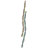
| Title | CRYSTAL STRUCTURE OF TROPOMYOSIN AT 7 ANGSTROMS RESOLUTION IN THE SPERMINE-INDUCED CRYSTAL FORM | ||||||
 Components Components | TROPOMYOSIN | ||||||
 Keywords Keywords | CONTRACTILE PROTEIN / TROPOMYOSIN COILED-COIL ALPHA-HELICAL | ||||||
| Function / homology |  Function and homology information Function and homology informationcardiac muscle contraction / actin filament organization / actin filament / actin filament binding / actin cytoskeleton / protein heterodimerization activity / protein homodimerization activity / identical protein binding / cytoplasm Similarity search - Function | ||||||
| Biological species |  | ||||||
| Method |  X-RAY DIFFRACTION / X-RAY DIFFRACTION /  SYNCHROTRON / SYNCHROTRON /  SIR / Resolution: 7 Å SIR / Resolution: 7 Å | ||||||
 Authors Authors | Whitby, F.G. / Phillips Jr., G.N. | ||||||
 Citation Citation |  Journal: Proteins / Year: 2000 Journal: Proteins / Year: 2000Title: Crystal structure of tropomyosin at 7 Angstroms resolution. Authors: Whitby, F.G. / Phillips Jr., G.N. | ||||||
| History |
|
- Structure visualization
| Structure viewer | Molecule:  Molmil Molmil Jmol/JSmol Jmol/JSmol |
|---|
- Downloads & links
- Download
| PDBx/mmCIF format |  1c1g.cif.gz 1c1g.cif.gz | 204 KB | Display |  PDBx/mmCIF format PDBx/mmCIF format |
|---|---|---|---|---|
| PDB format |  pdb1c1g.ent.gz pdb1c1g.ent.gz | 159.3 KB | Display |  PDB format PDB format |
| PDBx/mmJSON format |  1c1g.json.gz 1c1g.json.gz | Tree view |  PDBx/mmJSON format PDBx/mmJSON format | |
| Others |  Other downloads Other downloads |
-Validation report
| Summary document |  1c1g_validation.pdf.gz 1c1g_validation.pdf.gz | 460 KB | Display |  wwPDB validaton report wwPDB validaton report |
|---|---|---|---|---|
| Full document |  1c1g_full_validation.pdf.gz 1c1g_full_validation.pdf.gz | 614.1 KB | Display | |
| Data in XML |  1c1g_validation.xml.gz 1c1g_validation.xml.gz | 52.5 KB | Display | |
| Data in CIF |  1c1g_validation.cif.gz 1c1g_validation.cif.gz | 66.9 KB | Display | |
| Arichive directory |  https://data.pdbj.org/pub/pdb/validation_reports/c1/1c1g https://data.pdbj.org/pub/pdb/validation_reports/c1/1c1g ftp://data.pdbj.org/pub/pdb/validation_reports/c1/1c1g ftp://data.pdbj.org/pub/pdb/validation_reports/c1/1c1g | HTTPS FTP |
-Related structure data
| Similar structure data |
|---|
- Links
Links
- Assembly
| Deposited unit | 
| ||||||||
|---|---|---|---|---|---|---|---|---|---|
| 1 | 
| ||||||||
| 2 | 
| ||||||||
| Unit cell |
|
- Components
| #1: Protein | Mass: 32784.637 Da / Num. of mol.: 4 / Source method: isolated from a natural source / Source: (natural)  |
|---|
-Experimental details
-Experiment
| Experiment | Method:  X-RAY DIFFRACTION / Number of used crystals: 1 X-RAY DIFFRACTION / Number of used crystals: 1 |
|---|
- Sample preparation
| Crystal | Density Matthews: 3.7 Å3/Da / Density % sol: 68 % Description: Thimerosal was pre-reacted with purified tropomyosin prior growing crystals (spermine-induced crystal form). One mercury reacts with cyteine-190 of each chain of the molecule. Thus four ...Description: Thimerosal was pre-reacted with purified tropomyosin prior growing crystals (spermine-induced crystal form). One mercury reacts with cyteine-190 of each chain of the molecule. Thus four mercury atoms are present in the asymmetric unit. Two mercury atoms are located adjecent to each other on neighboring chains on the molecule, and for heavy-atom phasing purposes, these pairs were considered as a single point scatterer (due to 7 angstrom resolution limit). SIR phases determined from this derivative showed clear molecular packing consistent with the previously determined 9 angstrom structure in the same crystal form which was determined by molecular replacement using a highly simplified model (Whitby, et al. JMB 1992;227:441-452) | ||||||||||||||||||||
|---|---|---|---|---|---|---|---|---|---|---|---|---|---|---|---|---|---|---|---|---|---|
| Crystal grow | Temperature: 291 K / Method: liquid diffusion / pH: 7.4 Details: SPERMINE, pH 7.4, LIQUID DIFFUSION, temperature 18K | ||||||||||||||||||||
| Crystal grow | *PLUS Temperature: 15-17.5 ℃ | ||||||||||||||||||||
| Components of the solutions | *PLUS
|
-Data collection
| Diffraction | Mean temperature: 21 K | |||||||||
|---|---|---|---|---|---|---|---|---|---|---|
| Diffraction source | Source:  SYNCHROTRON / Site: SYNCHROTRON / Site:  NSLS NSLS  / Beamline: X8C / Wavelength: 1.000, 1.54 / Beamline: X8C / Wavelength: 1.000, 1.54 | |||||||||
| Detector | Detector: CCD | |||||||||
| Radiation | Protocol: SINGLE WAVELENGTH / Monochromatic (M) / Laue (L): M / Scattering type: x-ray | |||||||||
| Radiation wavelength |
| |||||||||
| Reflection | Resolution: 7→100 Å / Num. obs: 3117 / % possible obs: 96.38 % / Observed criterion σ(I): 0 / Redundancy: 3.4 % |
- Processing
| Software |
| ||||||||||||
|---|---|---|---|---|---|---|---|---|---|---|---|---|---|
| Refinement | Method to determine structure:  SIR / Resolution: 7→100 Å / σ(F): 0 SIR / Resolution: 7→100 Å / σ(F): 0
| ||||||||||||
| Refinement step | Cycle: LAST / Resolution: 7→100 Å
| ||||||||||||
| LS refinement shell | Resolution: 7→7.25 Å / Total num. of bins used: 10
|
 Movie
Movie Controller


 PDBj
PDBj