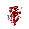
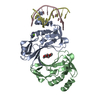
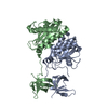
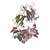
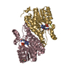
Mass: 26599.531 Da / Num. of mol.: 2 / Fragment: RESIDUES 49-286 / Mutation: F199K Source method: isolated from a genetically manipulated source Source: (gene. exp.) Rous sarcoma virus / Genus: Alpharetrovirus / Production host: Escherichia coli (E. coli) / References: UniProt: P03354, RNA-directed DNA polymerase
-
Experimental details
-
Experiment
Experiment
Method: X-RAY DIFFRACTION / Number of used crystals: 1
-
Sample preparation
Crystal
Density Matthews: 2.66 Å3/Da / Density % sol: 41.7 % Description: THE CATALYTIC DOMAIN WAS POSITIONED BY MOLECULAR REPLACEMENT USING AMORE WITH 1VSE AS STARTING MODEL. MAD PHASED MAP WAS CACULATED USING THE SAME ORIGIN AS THE MOLECULAR REPLACEMENT ...Description: THE CATALYTIC DOMAIN WAS POSITIONED BY MOLECULAR REPLACEMENT USING AMORE WITH 1VSE AS STARTING MODEL. MAD PHASED MAP WAS CACULATED USING THE SAME ORIGIN AS THE MOLECULAR REPLACEMENT SOLUTION AND SE POSITIONS WERE CONFIRMED. THE SH3- FOLD OF C-DOMAIN WAS LOCATED VISUALLY IN THE MAD MAP, HIV C-DOMAIN (1IHV) MONOMER WAS POSITIONED MANUALLY IN THE MAP AND MODIFIED TO RSV SEQUENCES DURING MODEL BUILDING.
Crystal grow
Details: ROD-SHAPED CRYSTALS WERE GROWN BY SITTING DROP VAPOR DIFFUSION METHOD. RESERVOIR SOLUTION CONTAINED 0.050M KCL, 0.01M MGCL2, 0.1M SODIUM CACODYLATE, 0.02M IMIDAZOLE, PH 7.2, 10% PEG3350, AND ...Details: ROD-SHAPED CRYSTALS WERE GROWN BY SITTING DROP VAPOR DIFFUSION METHOD. RESERVOIR SOLUTION CONTAINED 0.050M KCL, 0.01M MGCL2, 0.1M SODIUM CACODYLATE, 0.02M IMIDAZOLE, PH 7.2, 10% PEG3350, AND 20% (V/V) ETHYLENE GLYCOL. THE DROP CONTAINED 1:1 MIXTURE OF RESERVOIR SOLUTION AND PROTEIN (10-15 MG/ML)
Resolution: 3.1→15 Å / Cross valid method: THROUGHOUT / σ(F): 0.2 / Stereochemistry target values: ENGH & HUBER Details: THIS IS ONE OF SE-MET DATA SET COLLECTTED FOR MAD PHASING ALTHOUGH SE ATOMS WERE NOT MODELED. CAUTION SHOULD BE TAKEN IN INTERPRETING THOSE RESIDUES WITH B FACTORS IN UPPER 60S OR HIGHER
Rfactor
Num. reflection
% reflection
Selection details
Rfree
0.337
525
5.2 %
RANDOM
Rwork
0.256
-
-
-
obs
0.256
10183
5.1 %
-
Displacement parameters
Biso mean: 48 Å2
Baniso -1
Baniso -2
Baniso -3
1-
0.817 Å2
0 Å2
-10.779 Å2
2-
-
-3.077 Å2
0 Å2
3-
-
-
2.26 Å2
Refine analyze
Free
Obs
Luzzati coordinate error
0.58 Å
0.47 Å
Luzzati d res low
-
5 Å
Luzzati sigma a
0.7 Å
0.53 Å
Refinement step
Cycle: LAST / Resolution: 3.1→15 Å
Protein
Nucleic acid
Ligand
Solvent
Total
Num. atoms
3397
0
0
0
3397
Refine LS restraints
Refine-ID
Type
Dev ideal
X-RAY DIFFRACTION
c_bond_d
0.01
X-RAY DIFFRACTION
c_bond_d_na
X-RAY DIFFRACTION
c_bond_d_prot
X-RAY DIFFRACTION
c_angle_d
X-RAY DIFFRACTION
c_angle_d_na
X-RAY DIFFRACTION
c_angle_d_prot
X-RAY DIFFRACTION
c_angle_deg
1.41
X-RAY DIFFRACTION
c_angle_deg_na
X-RAY DIFFRACTION
c_angle_deg_prot
X-RAY DIFFRACTION
c_dihedral_angle_d
24
X-RAY DIFFRACTION
c_dihedral_angle_d_na
X-RAY DIFFRACTION
c_dihedral_angle_d_prot
X-RAY DIFFRACTION
c_improper_angle_d
0.97
X-RAY DIFFRACTION
c_improper_angle_d_na
X-RAY DIFFRACTION
c_improper_angle_d_prot
X-RAY DIFFRACTION
c_mcbond_it
X-RAY DIFFRACTION
c_mcangle_it
X-RAY DIFFRACTION
c_scbond_it
X-RAY DIFFRACTION
c_scangle_it
Refine LS restraints NCS
Ens-ID
Dom-ID
Refine-ID
Rms dev position (Å)
Weight position
1
1
X-RAY DIFFRACTION
0.062
200
2
2
X-RAY DIFFRACTION
0.097
150
LS refinement shell
Resolution: 3.1→3.21 Å
Rfactor
Num. reflection
% reflection
Rfree
0.459
42
4.15 %
Rwork
0.334
977
-
obs
-
-
96.5 %
Software
*PLUS
Name: 'CNS' / Classification: refinement
Refine LS restraints
*PLUS
Refine-ID
Type
Dev ideal
X-RAY DIFFRACTION
c_dihedral_angle_d
X-RAY DIFFRACTION
c_dihedral_angle_deg
24
X-RAY DIFFRACTION
c_improper_angle_d
X-RAY DIFFRACTION
c_improper_angle_deg
0.97
+
About Yorodumi
-
News
-
Feb 9, 2022. New format data for meta-information of EMDB entries
New format data for meta-information of EMDB entries
Version 3 of the EMDB header file is now the official format.
The previous official version 1.9 will be removed from the archive.
In the structure databanks used in Yorodumi, some data are registered as the other names, "COVID-19 virus" and "2019-nCoV". Here are the details of the virus and the list of structure data.
Jan 31, 2019. EMDB accession codes are about to change! (news from PDBe EMDB page)
EMDB accession codes are about to change! (news from PDBe EMDB page)
The allocation of 4 digits for EMDB accession codes will soon come to an end. Whilst these codes will remain in use, new EMDB accession codes will include an additional digit and will expand incrementally as the available range of codes is exhausted. The current 4-digit format prefixed with “EMD-” (i.e. EMD-XXXX) will advance to a 5-digit format (i.e. EMD-XXXXX), and so on. It is currently estimated that the 4-digit codes will be depleted around Spring 2019, at which point the 5-digit format will come into force.
The EM Navigator/Yorodumi systems omit the EMD- prefix.
Related info.:Q: What is EMD? / ID/Accession-code notation in Yorodumi/EM Navigator
Yorodumi is a browser for structure data from EMDB, PDB, SASBDB, etc.
This page is also the successor to EM Navigator detail page, and also detail information page/front-end page for Omokage search.
The word "yorodu" (or yorozu) is an old Japanese word meaning "ten thousand". "mi" (miru) is to see.
Related info.:EMDB / PDB / SASBDB / Comparison of 3 databanks / Yorodumi Search / Aug 31, 2016. New EM Navigator & Yorodumi / Yorodumi Papers / Jmol/JSmol / Function and homology information / Changes in new EM Navigator and Yorodumi
 Movie
Movie Controller


 Open data
 Basic information
 Components
 Keywords
 Function and homology information
 Rous sarcoma virus
 X-RAY DIFFRACTION /
  Authors
 Citation
 Structure visualization
 Downloads & links
 Other downloads


 PDBj
PDBj

 Assembly
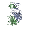

 Sample preparation
 / Beamline: X9B / Wavelength: 0.9791
  Processing