 Movie
Movie Controller

[English] 日本語
 Yorodumi
- PDB-1c0l: D-AMINO ACID OXIDASE: STRUCTURE OF SUBSTRATE COMPLEXES AT VERY HI... -
+ Open data

- Basic information
| Entry | Database: PDB / ID: 1c0l | ||||||
|---|---|---|---|---|---|---|---|
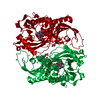
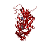
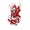
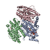
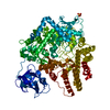
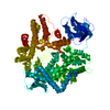
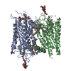
| Title | D-AMINO ACID OXIDASE: STRUCTURE OF SUBSTRATE COMPLEXES AT VERY HIGH RESOLUTION REVEAL THE CHEMICAL REACTTION MECHANISM OF FLAVIN DEHYDROGENATION | ||||||
 Components Components | D-AMINO ACID OXIDASE | ||||||
 Keywords Keywords | OXIDOREDUCTASE / FLAVIN CONTAINING PROTEIN ALPHA-BETA-ALPHA MOTIF | ||||||
| Function / homology |  Function and homology information Function and homology informationD-glutamate oxidase activity / D-amino acid metabolic process / D-amino-acid oxidase / D-amino-acid oxidase activity / glycine oxidase activity / D-amino acid catabolic process / nitrogen utilization / peroxisomal matrix / FAD binding / peroxisome Similarity search - Function | ||||||
| Biological species |  Rhodosporidium toruloides (fungus) Rhodosporidium toruloides (fungus) | ||||||
| Method |  X-RAY DIFFRACTION / Resolution: 1.73 Å X-RAY DIFFRACTION / Resolution: 1.73 Å | ||||||
 Authors Authors | Umhau, S. / Molla, G. / Diederichs, K. / Pilone, M.S. / Ghisla, S. / Welte, W. | ||||||
 Citation Citation | Journal: Proc.Natl.Acad.Sci.USA / Year: 2000 Title: The x-ray structure of D-amino acid oxidase at very high resolution identifies the chemical mechanism of flavin-dependent substrate dehydrogenation. Authors: Umhau, S. / Pollegioni, L. / Molla, G. / Diederichs, K. / Welte, W. / Pilone, M.S. / Ghisla, S. | ||||||
| History |
|
- Structure visualization
| Structure viewer | Molecule: MolmilJmol/JSmol |
|---|
- Downloads & links
-Download
| PDBx/mmCIF format | 1c0l.cif.gz | 99 KB | Display | PDBx/mmCIF format |
|---|---|---|---|---|
| PDB format | pdb1c0l.ent.gz | 74.5 KB | Display | PDB format |
| PDBx/mmJSON format | 1c0l.json.gz | Tree view | PDBx/mmJSON format | |
| Others |  Other downloads Other downloads |
-Validation report
| Arichive directory | https://data.pdbj.org/pub/pdb/validation_reports/c0/1c0lftp://data.pdbj.org/pub/pdb/validation_reports/c0/1c0l | HTTPS FTP |
|---|
-Related structure data


-Links
 PDBj
PDBj- Assembly
| Deposited unit | 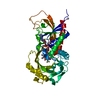
| |||||||||
|---|---|---|---|---|---|---|---|---|---|---|
| 1 | 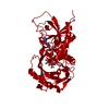
| |||||||||
| Unit cell |
| |||||||||
| Components on special symmetry positions |
|
-Components
| #1: Protein | Mass: 39614.922 Da / Num. of mol.: 1 Source method: isolated from a genetically manipulated source Source: (gene. exp.) Rhodosporidium toruloides (fungus) / Plasmid: PT7.7 / Production host:  |
|---|---|
| #2: Chemical | ChemComp-FLA /   Type: L-peptide linking / Mass: 143.065 Da / Num. of mol.: 1 / Source method: obtained synthetically / Formula: C3H4F3NO2 Type: L-peptide linking / Mass: 143.065 Da / Num. of mol.: 1 / Source method: obtained synthetically / Formula: C3H4F3NO2 |
| #3: Chemical | ChemComp-FAD /   Mass: 785.550 Da / Num. of mol.: 1 / Source method: obtained synthetically / Formula: C27H33N9O15P2 / Comment: FAD*YM Mass: 785.550 Da / Num. of mol.: 1 / Source method: obtained synthetically / Formula: C27H33N9O15P2 / Comment: FAD*YM |
| #4: Water | ChemComp-HOH /  Mass: 18.015 Da / Num. of mol.: 574 / Source method: isolated from a natural source / Formula: H2O Mass: 18.015 Da / Num. of mol.: 574 / Source method: isolated from a natural source / Formula: H2O |
-Experimental details
-Experiment
| Experiment | Method: X-RAY DIFFRACTION / Number of used crystals: 1 |
|---|
- Sample preparation
| Crystal | Density Matthews: 3.12 Å3/Da / Density % sol: 60.58 % | ||||||||||||||||||||||||||||||
|---|---|---|---|---|---|---|---|---|---|---|---|---|---|---|---|---|---|---|---|---|---|---|---|---|---|---|---|---|---|---|---|
| Crystal grow | Temperature: 289 K / Method: vapor diffusion, hanging drop / pH: 7.5 Details: 100MM HEPES 16% POLYETHYLENE GLYCOL PEG 200MM AMMONIUM SULFATE, pH 7.5, VAPOR DIFFUSION, HANGING DROP, temperature 289K | ||||||||||||||||||||||||||||||
| Crystal grow | *PLUS Temperature: 18 ℃ | ||||||||||||||||||||||||||||||
| Components of the solutions | *PLUS
|
-Data collection
| Diffraction source | Source: ROTATING ANODE / Type: OTHER / Wavelength: 1.5418 |
|---|---|
| Detector | Type: MARRESEARCH / Detector: IMAGE PLATE / Date: May 10, 1999 |
| Radiation | Protocol: SINGLE WAVELENGTH / Monochromatic (M) / Laue (L): M / Scattering type: x-ray |
| Radiation wavelength | Wavelength: 1.5418 Å / Relative weight: 1 |
| Reflection | Resolution: 1.7→100 Å / Num. all: 230642 / Num. obs: 52652 / % possible obs: 99.2 % / Observed criterion σ(F): 0 / Observed criterion σ(I): 0 / Redundancy: 4.4 % / Biso Wilson estimate: 23.7 Å2 / Rmerge(I) obs: 0.117 / Net I/σ(I): 6.4 |
| Reflection shell | Resolution: 1.73→1.79 Å / Redundancy: 1.8 % / Rmerge(I) obs: 0.282 / % possible all: 93.8 |
| Reflection | *PLUS Num. measured all: 230642 / Rmerge(I) obs: 0.11 |
| Reflection shell | *PLUS % possible obs: 96.7 % / Rmerge(I) obs: 0.372 / Mean I/σ(I) obs: 2.3 |
- Processing
| Software |
| |||||||||||||||||||||||||||||||||
|---|---|---|---|---|---|---|---|---|---|---|---|---|---|---|---|---|---|---|---|---|---|---|---|---|---|---|---|---|---|---|---|---|---|---|
| Refinement | Resolution: 1.73→100 Å / σ(F): 4 / σ(I): 4 / Stereochemistry target values: ENGH & HUBER
| |||||||||||||||||||||||||||||||||
| Refinement step | Cycle: LAST / Resolution: 1.73→100 Å
| |||||||||||||||||||||||||||||||||
| Refine LS restraints |
| |||||||||||||||||||||||||||||||||
| Software | *PLUS Name: SHELXL-97 / Classification: refinement | |||||||||||||||||||||||||||||||||
| Refinement | *PLUS Lowest resolution: 100 Å / σ(F): 4 / Rfactor Rwork: 0.162 | |||||||||||||||||||||||||||||||||
| Solvent computation | *PLUS | |||||||||||||||||||||||||||||||||
| Displacement parameters | *PLUS | |||||||||||||||||||||||||||||||||
| Refine LS restraints | *PLUS Type: s_angle_d / Dev ideal: 2.48 |
