 Movie
Movie Controller


+ Open data

- Basic information
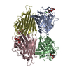
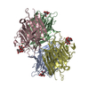
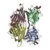
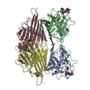
| Entry | Database: PDB / ID: 1bzw | |||||||||
|---|---|---|---|---|---|---|---|---|---|---|
| Title | PEANUT LECTIN COMPLEXED WITH C-LACTOSE | |||||||||
 Components Components | PROTEIN (PEANUT LECTIN) | |||||||||
 Keywords Keywords | SUGAR BINDING PROTEIN / LECTIN / LEGUME LECTIN / WATER BRIDGES / CARBOHYDRATE SPECIFICITY / C-LACTOSE / PROTEIN CRYSTALLOGRAPHY / AGGLUTININ | |||||||||
| Function / homology |  Function and homology information Function and homology information | |||||||||
| Biological species |  | |||||||||
| Method |  X-RAY DIFFRACTION / OTHER / Resolution: 2.7 Å X-RAY DIFFRACTION / OTHER / Resolution: 2.7 Å | |||||||||
 Authors Authors | Ravishankar, R. / Surolia, A. / Vijayan, M. / Lim, S. / Kishi, Y. | |||||||||
 Citation Citation | Journal: J.Am.Chem.Soc. / Year: 1998 Title: Preferred Conformation of C-Lactose at the Free and Peanut Lectin Bound States Authors: Ravishankar, R. / Surolia, A. / Vijayan, M. / Lim, S. / Kishi, Y. #1: Journal: Curr.Sci. / Year: 1997Title: The Specificity of Peanut Agglutinin for Thomsen-Friedenreich Antigen is Mediated by Water-Bridges Authors: Ravishankar, R. / Ravindran, M. / Suguna, K. / Surolia, A. / Vijayan, M. #2: Journal: J.Mol.Biol. / Year: 1996Title: Conformation, Protein-Carbohydrate Interactions and a Novel Subunit Association in the Refined Structure of Peanut Lectin-Lactose Complex Authors: Banerjee, R. / Das, K. / Ravishankar, R. / Suguna, K. / Surolia, A. / Vijayan, M. #3: Journal: J.Am.Chem.Soc. / Year: 1995Title: Biological Evaluation of Rationally Modified Analog of the H-Type II Blood Group Trisaccharide. A Correlation between Solution Conformation and Binding Affinity Authors: Wei, A. / Boy, K.M. / Kishi, Y. #4: Journal: J.Org.Chem. / Year: 1995Title: Preferred Conformation of C-Glycosides. 14. Synthes and Conformational Analysis of Carbon Analogs of Th Blood Group Determinant H-Type II Authors: Wei, A. / Haudrechy, A. / Audin, C. / Jun, H.-S. / Haudrechy-Brete, N. / Kishi, Y. #5: Journal: Proc.Natl.Acad.Sci.USA / Year: 1994Title: Crystal Structure of Peanut Lectin, a Protein with an Unusual Quaternary Structure Authors: Banerjee, R. / Mande, S.C. / Ganesh, V. / Das, K. / Dhanaraj, V. / Mahanta, S.K. / Suguna, K. / Surolia, A. / Vijayan, M. | |||||||||
| History |
|
- Structure visualization
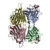
| Structure viewer | Molecule: MolmilJmol/JSmol |
|---|
- Downloads & links
-Download
| PDBx/mmCIF format | 1bzw.cif.gz | 192.4 KB | Display | PDBx/mmCIF format |
|---|---|---|---|---|
| PDB format | pdb1bzw.ent.gz | 154 KB | Display | PDB format |
| PDBx/mmJSON format | 1bzw.json.gz | Tree view | PDBx/mmJSON format | |
| Others |  Other downloads Other downloads |
-Validation report
| Arichive directory | https://data.pdbj.org/pub/pdb/validation_reports/bz/1bzwftp://data.pdbj.org/pub/pdb/validation_reports/bz/1bzw | HTTPS FTP |
|---|
-Related structure data
| Similar structure data |
|---|


-Links
 PDBj
PDBj


- Assembly
| Deposited unit | 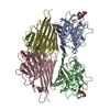
| ||||||||||||||||
|---|---|---|---|---|---|---|---|---|---|---|---|---|---|---|---|---|---|
| 1 |
| ||||||||||||||||
| Unit cell |
| ||||||||||||||||
| Noncrystallographic symmetry (NCS) | NCS oper:
|
-Components
-Protein , 1 types, 4 molecules ABCD
| #1: Protein | Mass: 24706.385 Da / Num. of mol.: 4 / Source method: isolated from a natural source / Source: (natural) |
|---|
-Sugars , 2 types, 4 molecules
| #2: Polysaccharide |   Source method: isolated from a genetically manipulated source Details: oligosaccharide / References: alpha-lactose #3: Polysaccharide | beta-D-galactopyranose-(1-4)-beta-D-glucopyranose / beta-lactose |   Source method: isolated from a genetically manipulated source Details: oligosaccharide / References: beta-lactose |
|---|
-Non-polymers , 3 types, 338 molecules 


| #4: Chemical | ChemComp-CA /  Mass: 40.078 Da / Num. of mol.: 4 / Source method: obtained synthetically / Formula: Ca Mass: 40.078 Da / Num. of mol.: 4 / Source method: obtained synthetically / Formula: Ca#5: Chemical | ChemComp-MN /  Mass: 54.938 Da / Num. of mol.: 4 / Source method: obtained synthetically / Formula: Mn Mass: 54.938 Da / Num. of mol.: 4 / Source method: obtained synthetically / Formula: Mn#6: Water | ChemComp-HOH / | Mass: 18.015 Da / Num. of mol.: 330 / Source method: isolated from a natural source / Formula: H2O |
|---|
-Experimental details
-Experiment
| Experiment | Method: X-RAY DIFFRACTION / Number of used crystals: 1 |
|---|
- Sample preparation
| Crystal | Density Matthews: 3.18 Å3/Da / Density % sol: 57 % | ||||||||||||||||||||||||||||||||||||||||||||||||||||||||||||||||||||||||
|---|---|---|---|---|---|---|---|---|---|---|---|---|---|---|---|---|---|---|---|---|---|---|---|---|---|---|---|---|---|---|---|---|---|---|---|---|---|---|---|---|---|---|---|---|---|---|---|---|---|---|---|---|---|---|---|---|---|---|---|---|---|---|---|---|---|---|---|---|---|---|---|---|---|
| Crystal grow | Method: vapor diffusion, hanging drop / pH: 7 Details: HANGING DROP OF 5 MG/ML PROTEIN IN 0.05 M SODIUM PHOSPHATE BUFFER,PH 7.0, CONTAINING 0.2M SODIUM CHLORIDE, 0.02 % SODIUM AZIDE, 10MM C-LACTOSE AND 12% (W/V) PEG 8000 IN THE SAME BUFFER, ...Details: HANGING DROP OF 5 MG/ML PROTEIN IN 0.05 M SODIUM PHOSPHATE BUFFER,PH 7.0, CONTAINING 0.2M SODIUM CHLORIDE, 0.02 % SODIUM AZIDE, 10MM C-LACTOSE AND 12% (W/V) PEG 8000 IN THE SAME BUFFER, vapor diffusion - hanging drop | ||||||||||||||||||||||||||||||||||||||||||||||||||||||||||||||||||||||||
| Crystal | *PLUS | ||||||||||||||||||||||||||||||||||||||||||||||||||||||||||||||||||||||||
| Crystal grow | *PLUS Method: vapor diffusion, hanging drop | ||||||||||||||||||||||||||||||||||||||||||||||||||||||||||||||||||||||||
| Components of the solutions | *PLUS
|
-Data collection
| Diffraction | Mean temperature: 293 K |
|---|---|
| Diffraction source | Source: ROTATING ANODE / Type: RIGAKU RU200 / Wavelength: 1.5418 |
| Detector | Type: MARRESEARCH / Detector: IMAGE PLATE / Details: MIRROR |
| Radiation | Protocol: SINGLE WAVELENGTH / Monochromatic (M) / Laue (L): M / Scattering type: x-ray |
| Radiation wavelength | Wavelength: 1.5418 Å / Relative weight: 1 |
| Reflection | Resolution: 2.7→19.7 Å / Num. obs: 33099 / % possible obs: 93.81 % / Redundancy: 3.75 % / Biso Wilson estimate: 30.8 Å2 / Rmerge(I) obs: 0.129 / Net I/σ(I): 14.2 |
| Reflection shell | Resolution: 2.7→2.8 Å / Redundancy: 2.9 % / Rmerge(I) obs: 0.461 / Mean I/σ(I) obs: 2.33 / % possible all: 93.8 |
| Reflection | *PLUS Num. measured all: 124179 |
- Processing
| Software |
| ||||||||||||||||||||||||||||||||||||||||||||||||||||||||||||
|---|---|---|---|---|---|---|---|---|---|---|---|---|---|---|---|---|---|---|---|---|---|---|---|---|---|---|---|---|---|---|---|---|---|---|---|---|---|---|---|---|---|---|---|---|---|---|---|---|---|---|---|---|---|---|---|---|---|---|---|---|---|
| Refinement | Method to determine structure: OTHER Starting model: PDB2PEL WITHOUT SUGAR AND WATER Resolution: 2.7→10 Å / Rfactor Rfree error: 0.007 / Data cutoff high absF: 10000000 / Data cutoff low absF: 0.001 / Isotropic thermal model: GROUP / Cross valid method: THROUGHOUT / σ(F): 0
| ||||||||||||||||||||||||||||||||||||||||||||||||||||||||||||
| Displacement parameters | Biso mean: 23.7 Å2
| ||||||||||||||||||||||||||||||||||||||||||||||||||||||||||||
| Refine analyze |
| ||||||||||||||||||||||||||||||||||||||||||||||||||||||||||||
| Refinement step | Cycle: LAST / Resolution: 2.7→10 Å
| ||||||||||||||||||||||||||||||||||||||||||||||||||||||||||||
| Refine LS restraints |
| ||||||||||||||||||||||||||||||||||||||||||||||||||||||||||||
| Refine LS restraints NCS | NCS model details: RESTRAIN | ||||||||||||||||||||||||||||||||||||||||||||||||||||||||||||
| LS refinement shell | Resolution: 2.7→2.87 Å / Rfactor Rfree error: 0.023 / Total num. of bins used: 6
| ||||||||||||||||||||||||||||||||||||||||||||||||||||||||||||
| Xplor file |
| ||||||||||||||||||||||||||||||||||||||||||||||||||||||||||||
| Software | *PLUS Name: X-PLOR / Version: 3.1 / Classification: refinement | ||||||||||||||||||||||||||||||||||||||||||||||||||||||||||||
| Refinement | *PLUS σ(F): 0 / % reflection Rfree: 3.9 % | ||||||||||||||||||||||||||||||||||||||||||||||||||||||||||||
| Solvent computation | *PLUS | ||||||||||||||||||||||||||||||||||||||||||||||||||||||||||||
| Displacement parameters | *PLUS Biso mean: 23.7 Å2 | ||||||||||||||||||||||||||||||||||||||||||||||||||||||||||||
| Refine LS restraints | *PLUS
| ||||||||||||||||||||||||||||||||||||||||||||||||||||||||||||
| LS refinement shell | *PLUS Rfactor Rfree: 0.264 / % reflection Rfree: 3.4 % / Rfactor Rwork: 0.27 |
