 Movie
Movie Controller

[English] 日本語
 Yorodumi
- PDB-1bw0: CRYSTAL STRUCTURE OF TYROSINE AMINOTRANSFERASE FROM TRYPANOSOMA CRUZI -
+ Open data

- Basic information
| Entry | Database: PDB / ID: 1bw0 | ||||||
|---|---|---|---|---|---|---|---|
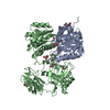
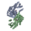
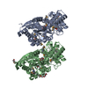
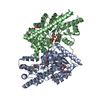
| Title | CRYSTAL STRUCTURE OF TYROSINE AMINOTRANSFERASE FROM TRYPANOSOMA CRUZI | ||||||
 Components Components | PROTEIN (TYROSINE AMINOTRANSFERASE) | ||||||
 Keywords Keywords | TRANSFERASE / TYROSINE CATABOLISM / AMINOTRANSFERASE / PYRIDOXAL-5'-PHOSPHATE / PLP | ||||||
| Function / homology |  Function and homology information Function and homology informationtyrosine transaminase / L-tyrosine:2-oxoglutarate transaminase activity / L-tyrosine catabolic process / L-phenylalanine catabolic process / pyridoxal phosphate binding / mitochondrion Similarity search - Function | ||||||
| Biological species |  | ||||||
| Method |  X-RAY DIFFRACTION / MOLECULAR REPLACEMENT / Resolution: 2.5 Å X-RAY DIFFRACTION / MOLECULAR REPLACEMENT / Resolution: 2.5 Å | ||||||
 Authors Authors | Blankenfeldt, W. / Montemartini, M. / Hunter, G.R. / Kalisz, H.M. / Nowicki, C. / Hecht, H.J. | ||||||
 Citation Citation | Journal: Protein Sci. / Year: 1999 Title: Crystal structure of Trypanosoma cruzi tyrosine aminotransferase: substrate specificity is influenced by cofactor binding mode. Authors: Blankenfeldt, W. / Nowicki, C. / Montemartini-Kalisz, M. / Kalisz, H.M. / Hecht, H.J. #1: Journal: Acta Crystallogr.,Sect.D / Year: 1998Title: Crystallization and Preliminary X-Ray Analysis of Tyrosine Aminotransferase from Trypanosoma Cruzi Epimastigotes Authors: Nowicki, C. / Montemartini, M. / Hunter, G.R. / Blankenfeldt, W. / Hecht, H.J. #2: Journal: Proc.Natl.Acad.Sci.USA / Year: 1996Title: Aromatic Amino Acid Transamination and Methionine Recycling in Trypanosomatids Authors: Berger, B.J. / Dai, W.W. / Wang, H. / Stark, R.E. / Cerami, A. #3: Journal: Fems Microbiol.Lett. / Year: 1994Title: Production of Aromatic Alpha-Hydroxyacids by Epimastigotes of Trypanosoma Cruzi, and its Possible Role in Nadh Reoxidation Authors: Montemartini, M. / Santome, J.A. / Cazzulo, J.J. / Nowicki, C. | ||||||
| History |
|
- Structure visualization
| Structure viewer | Molecule: MolmilJmol/JSmol |
|---|
- Downloads & links
-Download
| PDBx/mmCIF format | 1bw0.cif.gz | 170 KB | Display | PDBx/mmCIF format |
|---|---|---|---|---|
| PDB format | pdb1bw0.ent.gz | 134.1 KB | Display | PDB format |
| PDBx/mmJSON format | 1bw0.json.gz | Tree view | PDBx/mmJSON format | |
| Others |  Other downloads Other downloads |
-Validation report
| Arichive directory | https://data.pdbj.org/pub/pdb/validation_reports/bw/1bw0ftp://data.pdbj.org/pub/pdb/validation_reports/bw/1bw0 | HTTPS FTP |
|---|
-Related structure data
| Related structure data |  1artS S: Starting model for refinement |
|---|---|
| Similar structure data |


-Links
 PDBj
PDBj- Assembly
| Deposited unit | 
| ||||||||
|---|---|---|---|---|---|---|---|---|---|
| 1 |
| ||||||||
| Unit cell |
| ||||||||
| Noncrystallographic symmetry (NCS) | NCS oper: (Code: given Matrix: (0.57939, 0.761052, 0.291731), Vector: |
-Components
| #1: Protein | Mass: 46449.988 Da / Num. of mol.: 2 / Source method: isolated from a natural source Details: THE PROTEIN WAS PURIFIED FROM TRYPANOSOMA CRUZI EPIMASTIGOTES. Source: (natural) #2: Water | ChemComp-HOH / |  Mass: 18.015 Da / Num. of mol.: 118 / Source method: isolated from a natural source / Formula: H2O Mass: 18.015 Da / Num. of mol.: 118 / Source method: isolated from a natural source / Formula: H2O |
|---|
-Experimental details
-Experiment
| Experiment | Method: X-RAY DIFFRACTION / Number of used crystals: 5 |
|---|
- Sample preparation
| Crystal | Density Matthews: 2.5 Å3/Da / Density % sol: 51 % | ||||||||||||||||||||
|---|---|---|---|---|---|---|---|---|---|---|---|---|---|---|---|---|---|---|---|---|---|
| Crystal grow | pH: 7 Details: DIALYSIS OF 2.6 MG/ML PROTEIN AGAINST 25 % (W/W) PEG 8000, 5 MM PLP, 0.1 M PHOSPHATE/CITRATE, PH 7.0, 285 K | ||||||||||||||||||||
| Crystal | *PLUS | ||||||||||||||||||||
| Crystal grow | *PLUS Temperature: 285 K / Method: microdialysis | ||||||||||||||||||||
| Components of the solutions | *PLUS
|
-Data collection
| Diffraction | Mean temperature: 285 K |
|---|---|
| Diffraction source | Source: ROTATING ANODE / Type: SIEMENS / Wavelength: 1.5418 |
| Detector | Type: SIEMENS X1000 / Detector: AREA DETECTOR / Date: Jun 15, 1996 / Details: COLLIMATOR |
| Radiation | Protocol: SINGLE WAVELENGTH / Monochromatic (M) / Laue (L): M / Scattering type: x-ray |
| Radiation wavelength | Wavelength: 1.5418 Å / Relative weight: 1 |
| Reflection | Resolution: 2.5→10 Å / Num. obs: 24668 / % possible obs: 78.4 % / Redundancy: 1.7 % / Biso Wilson estimate: 30.17 Å2 / Rmerge(I) obs: 0.051 / Net I/σ(I): 17 |
| Reflection shell | Resolution: 2.5→2.64 Å / Redundancy: 1.2 % / Rmerge(I) obs: 0.096 / Mean I/σ(I) obs: 6 / % possible all: 47 |
| Reflection shell | *PLUS % possible obs: 47 % |
- Processing
| Software |
| ||||||||||||||||||||||||||||||||||||||||||||||||||||||||||||||||||||||||||||||||||||
|---|---|---|---|---|---|---|---|---|---|---|---|---|---|---|---|---|---|---|---|---|---|---|---|---|---|---|---|---|---|---|---|---|---|---|---|---|---|---|---|---|---|---|---|---|---|---|---|---|---|---|---|---|---|---|---|---|---|---|---|---|---|---|---|---|---|---|---|---|---|---|---|---|---|---|---|---|---|---|---|---|---|---|---|---|---|
| Refinement | Method to determine structure: MOLECULAR REPLACEMENT Starting model: PDB ENTRY 1ART Resolution: 2.5→10 Å / SU B: 5.63 / SU ML: 0.12 / Cross valid method: THROUGHOUT / σ(F): 0 / ESU R Free: 0.31
| ||||||||||||||||||||||||||||||||||||||||||||||||||||||||||||||||||||||||||||||||||||
| Displacement parameters | Biso mean: 25.19 Å2
| ||||||||||||||||||||||||||||||||||||||||||||||||||||||||||||||||||||||||||||||||||||
| Refinement step | Cycle: LAST / Resolution: 2.5→10 Å
| ||||||||||||||||||||||||||||||||||||||||||||||||||||||||||||||||||||||||||||||||||||
| Refine LS restraints |
| ||||||||||||||||||||||||||||||||||||||||||||||||||||||||||||||||||||||||||||||||||||
| Software | *PLUS Name: REFMAC / Classification: refinement | ||||||||||||||||||||||||||||||||||||||||||||||||||||||||||||||||||||||||||||||||||||
| Refinement | *PLUS Highest resolution: 2.5 Å / σ(F): 0 / % reflection Rfree: 5 % / Rfactor obs: 0.157 | ||||||||||||||||||||||||||||||||||||||||||||||||||||||||||||||||||||||||||||||||||||
| Solvent computation | *PLUS | ||||||||||||||||||||||||||||||||||||||||||||||||||||||||||||||||||||||||||||||||||||
| Displacement parameters | *PLUS | ||||||||||||||||||||||||||||||||||||||||||||||||||||||||||||||||||||||||||||||||||||
| Refine LS restraints | *PLUS
| ||||||||||||||||||||||||||||||||||||||||||||||||||||||||||||||||||||||||||||||||||||
| LS refinement shell | *PLUS Highest resolution: 2.51 Å / Lowest resolution: 2.6 Å / Rfactor Rfree: 0.294 / Rfactor obs: 0.211 |
