 Movie
Movie Controller

[English] 日本語
 Yorodumi
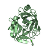
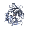
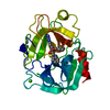
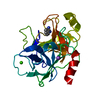
- PDB-1bit: THE CRYSTAL STRUCTURE OF ANIONIC SALMON TRYPSIN IN A SECOND CRYST... -
+ Open data

- Basic information
| Entry | Database: PDB / ID: 1bit | ||||||
|---|---|---|---|---|---|---|---|
| Title | THE CRYSTAL STRUCTURE OF ANIONIC SALMON TRYPSIN IN A SECOND CRYSTAL FORM | ||||||
 Components Components | TRYPSIN | ||||||
 Keywords Keywords | SERINE PROTEINASE | ||||||
| Function / homology |  Function and homology information Function and homology informationtrypsin / digestion / serine-type endopeptidase activity / proteolysis / : / metal ion binding Similarity search - Function | ||||||
| Biological species |  | ||||||
| Method |  X-RAY DIFFRACTION / Resolution: 1.83 Å X-RAY DIFFRACTION / Resolution: 1.83 Å | ||||||
 Authors Authors | Berglund, G.I. | ||||||
 Citation Citation | Journal: Acta Crystallogr.,Sect.D / Year: 1995 Title: Structure of anionic salmon trypsin in a second crystal form. Authors: Berglund, G.I. / Smalas, A.O. / Hordvik, A. / Willassen, N.P. #1: Journal: To be PublishedTitle: Cold-Adaption of Enzymes: Structural Comparison between Salmon and Bovine Trypsins Authors: Smalas, A.O. / Heimstad, E.S. / Hordvik, A. / Willassen, N.P. #2: Journal: Acta Crystallogr.,Sect.D / Year: 1993Title: Crystal Structure Determination and Refinement of Benzamidine-Inhibited Trypsin from the North Atlantic Salmon (Salmo Salar) Authors: Smalas, A.O. / Hordvik, A. #3: Journal: J.Mol.Biol. / Year: 1990Title: Crystallization and Preliminary X-Ray Crystallographic Studies of Benzamidine-Inhibited Trypsin from the North Atlantic Salmon (Salmo Salar) Authors: Smalas, A.O. / Hordvik, A. / Hansen, L.K. / Hough, E. / Jynge, K. #4: Journal: J.Mol.Biol. / Year: 1989Title: Crystal Structure of Bovine B-Trypsin at 1.5 Angstroms Resolution in a Crystal Form with Low Molecular Packing Density Authors: Bartunik, H.D. / Summers, L.J. / Bartsch, H.H. #5: Journal: Acta Crystallogr.,Sect.B / Year: 1983Title: The Geometry of the Reactive Site and of the Peptide Groups in Trypsin, Trypsinogen and its Complexes with Inhibitors Authors: Marquart, M. / Walter, J. / Deisenhofer, J. / Bode, W. / Huber, R. #6: Journal: Acta Crystallogr.,Sect.B / Year: 1979Title: The Accuracy of Refined Protein Structures: Comparison of Two Independently Refined Models of Bovine Trypsin Authors: Chambers, J.H. / Stroud, R.M. | ||||||
| History |
|
- Structure visualization
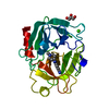
| Structure viewer | Molecule: MolmilJmol/JSmol |
|---|
- Downloads & links
-Download
| PDBx/mmCIF format | 1bit.cif.gz | 59.6 KB | Display | PDBx/mmCIF format |
|---|---|---|---|---|
| PDB format | pdb1bit.ent.gz | 42.9 KB | Display | PDB format |
| PDBx/mmJSON format | 1bit.json.gz | Tree view | PDBx/mmJSON format | |
| Others |  Other downloads Other downloads |
-Validation report
| Arichive directory | https://data.pdbj.org/pub/pdb/validation_reports/bi/1bitftp://data.pdbj.org/pub/pdb/validation_reports/bi/1bit | HTTPS FTP |
|---|
-Related structure data
| Similar structure data |
|---|


-Links
 PDBj
PDBj


- Assembly
| Deposited unit | 
| ||||||||
|---|---|---|---|---|---|---|---|---|---|
| 1 |
| ||||||||
| Unit cell |
|
-Components
| #1: Protein | Mass: 25402.500 Da / Num. of mol.: 1 Source method: isolated from a genetically manipulated source Source: (gene. exp.) | ||||||||
|---|---|---|---|---|---|---|---|---|---|
| #2: Chemical | ChemComp-CA /   Mass: 40.078 Da / Num. of mol.: 1 / Source method: obtained synthetically / Formula: Ca Mass: 40.078 Da / Num. of mol.: 1 / Source method: obtained synthetically / Formula: Ca | ||||||||
| #3: Chemical | ChemComp-SO4 /   Mass: 96.063 Da / Num. of mol.: 1 / Source method: obtained synthetically / Formula: SO4 Mass: 96.063 Da / Num. of mol.: 1 / Source method: obtained synthetically / Formula: SO4 | ||||||||
| #4: Chemical |   Mass: 120.152 Da / Num. of mol.: 2 / Source method: obtained synthetically / Formula: C7H8N2 Mass: 120.152 Da / Num. of mol.: 2 / Source method: obtained synthetically / Formula: C7H8N2#5: Water | ChemComp-HOH / |  Mass: 18.015 Da / Num. of mol.: 125 / Source method: isolated from a natural source / Formula: H2O Mass: 18.015 Da / Num. of mol.: 125 / Source method: isolated from a natural source / Formula: H2OHas protein modification | Y | Nonpolymer details | CALCIUM (IDENTIFIER CA) IS STRUCTURALLY BOUND IN THE SAME SEQUENTIAL POSITION AS FOR BOVINE TRYPSIN. ...CALCIUM (IDENTIFIER | Sequence details | THE AMINO ACID NUMBERING SYSTEM IS THE ONE ADOPTED FROM CHYMOTRYPSINOGEN AND IS ALSO THE SAME AS ...THE AMINO ACID NUMBERING SYSTEM IS THE ONE ADOPTED FROM CHYMOTRYPS | |
-Experimental details
-Experiment
| Experiment | Method: X-RAY DIFFRACTION |
|---|
- Sample preparation
| Crystal | Density Matthews: 1.95 Å3/Da / Density % sol: 36.78 % | ||||||||||||||||||||||||||||||||||||
|---|---|---|---|---|---|---|---|---|---|---|---|---|---|---|---|---|---|---|---|---|---|---|---|---|---|---|---|---|---|---|---|---|---|---|---|---|---|
| Crystal grow | *PLUS pH: 6 / Method: vapor diffusion | ||||||||||||||||||||||||||||||||||||
| Components of the solutions | *PLUS
|
-Data collection
| Radiation | Scattering type: x-ray |
|---|---|
| Radiation wavelength | Relative weight: 1 |
| Reflection | *PLUS Highest resolution: 1.83 Å / Num. obs: 15984 / % possible obs: 88.2 % / Num. measured all: 25917 / Rmerge F obs: 0.06 |
- Processing
| Software |
| ||||||||||||||||||||||||||||||||||||||||||||||||||||||||||||
|---|---|---|---|---|---|---|---|---|---|---|---|---|---|---|---|---|---|---|---|---|---|---|---|---|---|---|---|---|---|---|---|---|---|---|---|---|---|---|---|---|---|---|---|---|---|---|---|---|---|---|---|---|---|---|---|---|---|---|---|---|---|
| Refinement | Resolution: 1.83→8 Å / σ(F): 3 /
| ||||||||||||||||||||||||||||||||||||||||||||||||||||||||||||
| Refinement step | Cycle: LAST / Resolution: 1.83→8 Å
| ||||||||||||||||||||||||||||||||||||||||||||||||||||||||||||
| Refine LS restraints |
| ||||||||||||||||||||||||||||||||||||||||||||||||||||||||||||
| Software | *PLUS Name: X-PLOR/PROLSQ / Classification: refinement | ||||||||||||||||||||||||||||||||||||||||||||||||||||||||||||
| Refinement | *PLUS Rfactor obs: 0.199 / Rfactor Rfree: 0.272 | ||||||||||||||||||||||||||||||||||||||||||||||||||||||||||||
| Solvent computation | *PLUS | ||||||||||||||||||||||||||||||||||||||||||||||||||||||||||||
| Displacement parameters | *PLUS Biso mean: 14.2 Å2 | ||||||||||||||||||||||||||||||||||||||||||||||||||||||||||||
| Refine LS restraints | *PLUS
|
