 Movie
Movie Controller

[English] 日本語
 Yorodumi
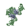
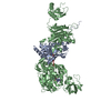
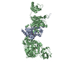
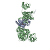
- PDB-1b7y: PHENYLALANYL TRNA SYNTHETASE COMPLEXED WITH PHENYLALANINYL-ADENYLATE -
+ Open data

- Basic information
| Entry | Database: PDB / ID: 1b7y | ||||||
|---|---|---|---|---|---|---|---|
| Title | PHENYLALANYL TRNA SYNTHETASE COMPLEXED WITH PHENYLALANINYL-ADENYLATE | ||||||
 Components Components | (PROTEIN (PHENYLALANYL-TRNA SYNTHETASE)) x 2 | ||||||
 Keywords Keywords | LIGASE / ENZYME / TRNA SYNTHETASE / ALPHA/BETA HOMODIMER | ||||||
| Function / homology |  Function and homology information Function and homology informationphenylalanine-tRNA ligase complex / phenylalanine-tRNA ligase / phenylalanyl-tRNA aminoacylation / phenylalanine-tRNA ligase activity / tRNA binding / magnesium ion binding / ATP binding / cytoplasm Similarity search - Function | ||||||
| Biological species |   Thermus thermophilus (bacteria) Thermus thermophilus (bacteria) | ||||||
| Method |  X-RAY DIFFRACTION / SYNCHROTRON / MOLECULAR REPLACEMENT / Resolution: 2.5 Å X-RAY DIFFRACTION / SYNCHROTRON / MOLECULAR REPLACEMENT / Resolution: 2.5 Å | ||||||
 Authors Authors | Reshetnikova, L. / Moor, N. / Lavrik, O. / Vassylyev, D.G. | ||||||
 Citation Citation | Journal: J.Mol.Biol. / Year: 1999 Title: Crystal structures of phenylalanyl-tRNA synthetase complexed with phenylalanine and a phenylalanyl-adenylate analogue. Authors: Reshetnikova, L. / Moor, N. / Lavrik, O. / Vassylyev, D.G. #1: Journal: Structure / Year: 1997Title: The crystal structure of phenylalanyl-tRNA synthetase from thermus thermophilus complexed with cognate tRNAPhe. Authors: Goldgur, Y. / Mosyak, L. / Reshetnikova, L. / Ankilova, V. / Lavrik, O. / Khodyreva, S. / Safro, M. #2: Journal: Nature New Biol. / Year: 1995Title: Structure of Phenylalanyl-tRNA Synthetase from Thermus Thermophilus Authors: Mosyak, L. / Reshetnikova, L. / Goldgur, Y. / Delarue, M. / Safro, M.G. | ||||||
| History |
|
- Structure visualization
| Structure viewer | Molecule: MolmilJmol/JSmol |
|---|
- Downloads & links
-Download
| PDBx/mmCIF format | 1b7y.cif.gz | 224.7 KB | Display | PDBx/mmCIF format |
|---|---|---|---|---|
| PDB format | pdb1b7y.ent.gz | 175.5 KB | Display | PDB format |
| PDBx/mmJSON format | 1b7y.json.gz | Tree view | PDBx/mmJSON format | |
| Others |  Other downloads Other downloads |
-Validation report
| Arichive directory | https://data.pdbj.org/pub/pdb/validation_reports/b7/1b7yftp://data.pdbj.org/pub/pdb/validation_reports/b7/1b7y | HTTPS FTP |
|---|
-Related structure data
| Related structure data | 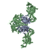 1b70C 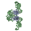 1pysS S: Starting model for refinement C: citing same article ( |
|---|---|
| Similar structure data |


-Links
 PDBj
PDBj

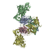
- Assembly
| Deposited unit | 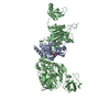
| ||||||||||
|---|---|---|---|---|---|---|---|---|---|---|---|
| 1 | 
| ||||||||||
| Unit cell |
|
-Components
| #1: Protein | Mass: 39309.199 Da / Num. of mol.: 1 / Source method: isolated from a natural source / Source: (natural) Thermus thermophilus (bacteria) / Cellular location: CYTOPLASM / References: UniProt: P27001, phenylalanine-tRNA ligase |
|---|---|
| #2: Protein | Mass: 86720.656 Da / Num. of mol.: 1 / Source method: isolated from a natural source / Source: (natural) Thermus thermophilus (bacteria) / Cellular location: CYTOPLASM / References: UniProt: P27002, phenylalanine-tRNA ligase |
| #3: Chemical | ChemComp-MG /   Mass: 24.305 Da / Num. of mol.: 1 / Source method: obtained synthetically / Formula: Mg Mass: 24.305 Da / Num. of mol.: 1 / Source method: obtained synthetically / Formula: Mg |
| #4: Chemical | ChemComp-FYA / 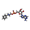  Mass: 480.412 Da / Num. of mol.: 1 / Source method: obtained synthetically / Formula: C19H25N6O7P Mass: 480.412 Da / Num. of mol.: 1 / Source method: obtained synthetically / Formula: C19H25N6O7P |
| #5: Water | ChemComp-HOH /  Mass: 18.015 Da / Num. of mol.: 239 / Source method: isolated from a natural source / Formula: H2O Mass: 18.015 Da / Num. of mol.: 239 / Source method: isolated from a natural source / Formula: H2O |
-Experimental details
-Experiment
| Experiment | Method: X-RAY DIFFRACTION / Number of used crystals: 1 |
|---|
- Sample preparation
| Crystal | Density Matthews: 3.8 Å3/Da / Density % sol: 70 % | ||||||||||||||||||||||||||||||||||||||||||||||||
|---|---|---|---|---|---|---|---|---|---|---|---|---|---|---|---|---|---|---|---|---|---|---|---|---|---|---|---|---|---|---|---|---|---|---|---|---|---|---|---|---|---|---|---|---|---|---|---|---|---|
| Crystal grow | Temperature: 313 K / Method: vapor diffusion, hanging drop / pH: 7 Details: HANGING-DROP TECHNIQUE AT 4O C WITH (NH4)2SO4 AS PRECIPITANT. EACH DROPLET OF THE PHENYLALANYL-TRNA SYNTHETASE SOLUTION (10 microliters) AT PROTEIN CONCENTRATION OF 3 TO 5 MG PER ML IN 20 MM ...Details: HANGING-DROP TECHNIQUE AT 4O C WITH (NH4)2SO4 AS PRECIPITANT. EACH DROPLET OF THE PHENYLALANYL-TRNA SYNTHETASE SOLUTION (10 microliters) AT PROTEIN CONCENTRATION OF 3 TO 5 MG PER ML IN 20 MM IMIDASOL-HCL BUFFER (PH 7.8), 1 MM MGCL2, 1 MM NAN3 AND 15% SATURATED (NH4)2SO4 IN THE SAME BUFFER. CRYSTALS APPEARED AFTER ONE TO TWO WEEKS AND GREW UP TO THE MAXIMAL DIMENSIONS OF 0.4X0.4X0.25 MM IN A FEW WEEKS, pH 7.0, VAPOR DIFFUSION, HANGING DROP, temperature 313K | ||||||||||||||||||||||||||||||||||||||||||||||||
| Components of the solutions |
| ||||||||||||||||||||||||||||||||||||||||||||||||
| Crystal | *PLUS Density % sol: 75 % | ||||||||||||||||||||||||||||||||||||||||||||||||
| Crystal grow | *PLUS Temperature: 4 ℃ / pH: 7.8 / Details: Chernaya, M., (1987) J. Mol. Biol., 198, 555. | ||||||||||||||||||||||||||||||||||||||||||||||||
| Components of the solutions | *PLUS
|
-Data collection
| Diffraction | Mean temperature: 100 K |
|---|---|
| Diffraction source | Source: SYNCHROTRON / Site: SPring-8  / Beamline: BL41XU / Wavelength: 1 / Beamline: BL41XU / Wavelength: 1 |
| Detector | Type: RIGAKU / Detector: IMAGE PLATE / Date: Dec 15, 1998 |
| Radiation | Protocol: SINGLE WAVELENGTH / Monochromatic (M) / Laue (L): M / Scattering type: x-ray |
| Radiation wavelength | Wavelength: 1 Å / Relative weight: 1 |
| Reflection | Resolution: 2.5→50 Å / Num. all: 81613 / Num. obs: 81613 / % possible obs: 95.7 % / Observed criterion σ(I): 0 / Redundancy: 4 % / Biso Wilson estimate: 45 Å2 / Rmerge(I) obs: 0.064 / Net I/σ(I): 6 |
| Reflection shell | Resolution: 2.5→2.59 Å / Redundancy: 4 % / Rmerge(I) obs: 0.399 / Mean I/σ(I) obs: 2.2 / % possible all: 88.5 |
| Reflection | *PLUS |
| Reflection shell | *PLUS % possible obs: 88.5 % |
- Processing
| Software |
| ||||||||||||||||||||||||||||||||||||||||||||||||||||||||||||||||||||||||||||||||
|---|---|---|---|---|---|---|---|---|---|---|---|---|---|---|---|---|---|---|---|---|---|---|---|---|---|---|---|---|---|---|---|---|---|---|---|---|---|---|---|---|---|---|---|---|---|---|---|---|---|---|---|---|---|---|---|---|---|---|---|---|---|---|---|---|---|---|---|---|---|---|---|---|---|---|---|---|---|---|---|---|---|
| Refinement | Method to determine structure: MOLECULAR REPLACEMENT Starting model: PDB ENTRY 1PYS Resolution: 2.5→50 Å / Data cutoff high absF: 0 / Data cutoff low absF: 0 / Cross valid method: THROUGHOUT / σ(F): 0
| ||||||||||||||||||||||||||||||||||||||||||||||||||||||||||||||||||||||||||||||||
| Displacement parameters | Biso mean: 62 Å2 | ||||||||||||||||||||||||||||||||||||||||||||||||||||||||||||||||||||||||||||||||
| Refine analyze | Luzzati coordinate error obs: 0.25 Å / Luzzati d res low obs: 5 Å / Luzzati sigma a obs: 0.22 Å | ||||||||||||||||||||||||||||||||||||||||||||||||||||||||||||||||||||||||||||||||
| Refinement step | Cycle: LAST / Resolution: 2.5→50 Å
| ||||||||||||||||||||||||||||||||||||||||||||||||||||||||||||||||||||||||||||||||
| Refine LS restraints |
| ||||||||||||||||||||||||||||||||||||||||||||||||||||||||||||||||||||||||||||||||
| LS refinement shell | Resolution: 2.5→2.61 Å / Total num. of bins used: 8
| ||||||||||||||||||||||||||||||||||||||||||||||||||||||||||||||||||||||||||||||||
| Xplor file |
| ||||||||||||||||||||||||||||||||||||||||||||||||||||||||||||||||||||||||||||||||
| Software | *PLUS Name: X-PLOR / Version: 3.851 / Classification: refinement | ||||||||||||||||||||||||||||||||||||||||||||||||||||||||||||||||||||||||||||||||
| Refine LS restraints | *PLUS
|
