| 登録情報 | データベース: PDB / ID: 1b4f
|
|---|
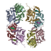
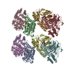
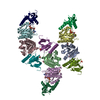
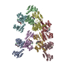
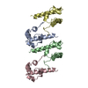
| タイトル | OLIGOMERIC STRUCTURE OF THE HUMAN EPHB2 RECEPTOR SAM DOMAIN |
|---|
 要素 要素 | EPHB2 |
|---|
 キーワード キーワード | SIGNAL TRANSDUCTION / SAM DOMAIN / EPH RECEPTOR / OLIGOMER |
|---|
| 機能・相同性 |  機能・相同性情報 機能・相同性情報
regulation of T-helper 17 type immune response / hindbrain tangential cell migration / negative regulation of glutamate receptor signaling pathway / vesicle-mediated intercellular transport / L1CAM interactions / optic nerve morphogenesis / urogenital system development / tight junction assembly / regulation of body fluid levels / neuron projection retraction ...regulation of T-helper 17 type immune response / hindbrain tangential cell migration / negative regulation of glutamate receptor signaling pathway / vesicle-mediated intercellular transport / L1CAM interactions / optic nerve morphogenesis / urogenital system development / tight junction assembly / regulation of body fluid levels / neuron projection retraction / central nervous system projection neuron axonogenesis / postsynaptic membrane assembly / axon guidance receptor activity / transmembrane-ephrin receptor activity / negative regulation of axonogenesis / positive regulation of synaptic plasticity / positive regulation of long-term neuronal synaptic plasticity / corpus callosum development / dendritic spine development / camera-type eye morphogenesis / regulation of filopodium assembly / positive regulation of dendritic spine morphogenesis / positive regulation of glutamate receptor signaling pathway / regulation of autophagosome assembly / regulation of behavioral fear response / commissural neuron axon guidance / dendritic spine morphogenesis / negative regulation of cell adhesion / negative regulation of Ras protein signal transduction / positive regulation of protein localization to cell surface / phosphorylation / axonal fasciculation / retinal ganglion cell axon guidance / EPH-Ephrin signaling / Ephrin signaling / positive regulation of synapse assembly / regulation of receptor signaling pathway via JAK-STAT / positive regulation of immunoglobulin production / inner ear morphogenesis / regulation of blood coagulation / B cell activation / roof of mouth development / EPH-ephrin mediated repulsion of cells / regulation of neuronal synaptic plasticity / ephrin receptor signaling pathway / positive regulation of B cell proliferation / EPHB-mediated forward signaling / neuron projection maintenance / negative regulation of cytokine production involved in inflammatory response / axon guidance / peptidyl-tyrosine phosphorylation / hippocampal mossy fiber to CA3 synapse / positive regulation of long-term synaptic potentiation / learning / positive regulation of protein localization to plasma membrane / receptor protein-tyrosine kinase / negative regulation of ERK1 and ERK2 cascade / cellular response to amyloid-beta / positive regulation of tumor necrosis factor production / nervous system development / amyloid-beta binding / cellular response to lipopolysaccharide / presynaptic membrane / protein tyrosine kinase activity / angiogenesis / dendritic spine / postsynaptic membrane / learning or memory / postsynapse / positive regulation of cell migration / signaling receptor binding / axon / neuronal cell body / dendrite / positive regulation of gene expression / protein-containing complex binding / glutamatergic synapse / cell surface / extracellular region / nucleoplasm / ATP binding / identical protein binding / plasma membrane / cytosol類似検索 - 分子機能 Ephrin type-B receptor 2, ligand binding domain / Transcription Factor, Ets-1 / Tyrosine-protein kinase ephrin type A/B receptor-like / Tyrosine-protein kinase ephrin type A/B receptor-like / Ephrin receptor type-A /type-B / Ephrin receptor ligand binding domain / Tyrosine-protein kinase, receptor class V, conserved site / Ephrin receptor, transmembrane domain / : / Ephrin receptor ligand binding domain ...Ephrin type-B receptor 2, ligand binding domain / Transcription Factor, Ets-1 / Tyrosine-protein kinase ephrin type A/B receptor-like / Tyrosine-protein kinase ephrin type A/B receptor-like / Ephrin receptor type-A /type-B / Ephrin receptor ligand binding domain / Tyrosine-protein kinase, receptor class V, conserved site / Ephrin receptor, transmembrane domain / : / Ephrin receptor ligand binding domain / Ephrin type-A receptor 2 transmembrane domain / Receptor tyrosine kinase class V signature 1. / Receptor tyrosine kinase class V signature 2. / Eph receptor ligand-binding domain profile. / Ephrin receptor ligand binding domain / Putative ephrin-receptor like / SAM domain (Sterile alpha motif) / SAM domain profile. / Sterile alpha motif. / Sterile alpha motif domain / Sterile alpha motif/pointed domain superfamily / Growth factor receptor cysteine-rich domain superfamily / Fibronectin type III domain / Fibronectin type 3 domain / Fibronectin type-III domain profile. / Galactose-binding-like domain superfamily / Fibronectin type III / Fibronectin type III superfamily / DNA polymerase; domain 1 / Tyrosine-protein kinase, catalytic domain / Tyrosine kinase, catalytic domain / Tyrosine protein kinases specific active-site signature. / Tyrosine-protein kinase, active site / Serine-threonine/tyrosine-protein kinase, catalytic domain / Protein tyrosine and serine/threonine kinase / Protein kinase, ATP binding site / Protein kinases ATP-binding region signature. / Immunoglobulin-like fold / Protein kinase domain profile. / Protein kinase domain / Protein kinase-like domain superfamily / Orthogonal Bundle / Mainly Alpha類似検索 - ドメイン・相同性 |
|---|
| 生物種 |  Homo sapiens (ヒト) Homo sapiens (ヒト) |
|---|
| 手法 |  X線回折 / シンクロトロン / 多波長異常分散 / 解像度: 1.95 Å X線回折 / シンクロトロン / 多波長異常分散 / 解像度: 1.95 Å |
|---|
 データ登録者 データ登録者 | Thanos, C.D. / Goodwill, K.E. / Bowie, J.U. |
|---|
 引用 引用 | ジャーナル: Science / 年: 1999
タイトル: Oligomeric structure of the human EphB2 receptor SAM domain.
著者: Thanos, C.D. / Goodwill, K.E. / Bowie, J.U. |
|---|
| 履歴 | | 登録 | 1998年12月20日 | 処理サイト: BNL |
|---|
| 改定 1.0 | 1999年2月16日 | Provider: repository / タイプ: Initial release |
|---|
| 改定 1.1 | 2008年3月24日 | Group: Version format compliance |
|---|
| 改定 1.2 | 2011年7月13日 | Group: Version format compliance |
|---|
| 改定 1.3 | 2019年12月11日 | Group: Advisory / Database references / Derived calculations
カテゴリ: pdbx_distant_solvent_atoms / pdbx_struct_assembly ...pdbx_distant_solvent_atoms / pdbx_struct_assembly / pdbx_struct_assembly_gen / pdbx_struct_oper_list / struct_ref_seq_dif
Item: _struct_ref_seq_dif.details |
|---|
| 改定 1.4 | 2024年2月7日 | Group: Data collection / Database references / カテゴリ: chem_comp_atom / chem_comp_bond / database_2
Item: _database_2.pdbx_DOI / _database_2.pdbx_database_accession |
|---|
|
|---|
 ムービー
ムービー コントローラー


 データを開く
 基本情報
基本情報 構造の表示
構造の表示 ダウンロードとリンク
 その他のダウンロード
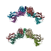
 PDBj
PDBj


 集合体
集合体


 分子量: 18.015 Da / 分子数: 685 / 由来タイプ: 天然 / 式: H2O
 試料調製
試料調製 / ビームライン: 5.0.2 / 波長: 0.96859, 0.97966, 0.97982, 1.0000
 解析
解析