 Movie
Movie Controller


+ Open data

- Basic information
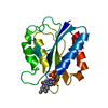
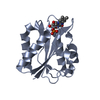
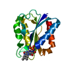
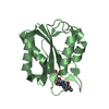
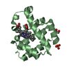
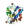
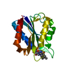
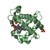
| Entry | Database: PDB / ID: 1azl | ||||||
|---|---|---|---|---|---|---|---|
| Title | G61V FLAVODOXIN MUTANT FROM DESULFOVIBRIO VULGARIS | ||||||
 Components Components | FLAVODOXIN | ||||||
 Keywords Keywords | ELECTRON TRANSPORT / ELECTRON TRANSFER / FLAVOPROTEIN / FMN / FLAVODOXIN / MUTANT | ||||||
| Function / homology |  Function and homology information Function and homology information | ||||||
| Biological species |  Desulfovibrio vulgaris subsp. vulgaris str. Hildenborough (bacteria) Desulfovibrio vulgaris subsp. vulgaris str. Hildenborough (bacteria) | ||||||
| Method |  X-RAY DIFFRACTION / SYNCHROTRON / MOLECULAR REPLACEMENT / Resolution: 1.8 Å X-RAY DIFFRACTION / SYNCHROTRON / MOLECULAR REPLACEMENT / Resolution: 1.8 Å | ||||||
 Authors Authors | Walsh, M.A. / Mccarthy, A. / O'Farrell, P.A. / Voordouw, G. / Higgins, T. / Mayhew, S.G. | ||||||
 Citation Citation | Journal: Biochemistry / Year: 1998 Title: Modulation of the redox potentials of FMN in Desulfovibrio vulgaris flavodoxin: thermodynamic properties and crystal structures of glycine-61 mutants. Authors: O'Farrell, P.A. / Walsh, M.A. / McCarthy, A.A. / Higgins, T.M. / Voordouw, G. / Mayhew, S.G. #1: Journal: J.Mol.Biol. / Year: 1991Title: Comparison of the Crystal Structures of a Flavodoxin in its Three Oxidation States at Cryogenic Temperatures Authors: Watt, W. / Tulinsky, A. / Swenson, R.P. / Watenpaugh, K.D. #2: Journal: J.Biol.Chem. / Year: 1988Title: Cloning,Nucleotide Sequence,and Expression of the Flavodoxin Gene from Desulfovibrio Vulgaris (Hildenborough) Authors: Krey, G.D. / Vanin, E.F. / Swenson, R.P. #3: Journal: Fems Microbiol.Lett. / Year: 1988Title: Cloning and Sequencing of the Gene Encoding Flavodoxin from Desulfovibrio Vulgaris Hildenborough Authors: Curley, G.P. / Voordouw, G. #4: Journal: Proc.Natl.Acad.Sci.USA / Year: 1972Title: Structure of the Oxidized Form of a Flavodoxin at 2.5-Angstrom Resolution:Resolution of the Phase Ambiguity by Anomalous Scattering Authors: Watenpaugh, K.D. / Sieker, L.C. / Jensen, L.H. / Legall, J. / Dubourdieu, M. | ||||||
| History |
|
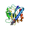
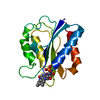
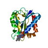
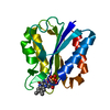
- Structure visualization
| Structure viewer | Molecule: MolmilJmol/JSmol |
|---|
- Downloads & links
-Download
| PDBx/mmCIF format | 1azl.cif.gz | 45.2 KB | Display | PDBx/mmCIF format |
|---|---|---|---|---|
| PDB format | pdb1azl.ent.gz | 30.9 KB | Display | PDB format |
| PDBx/mmJSON format | 1azl.json.gz | Tree view | PDBx/mmJSON format | |
| Others |  Other downloads Other downloads |
-Validation report
| Arichive directory | https://data.pdbj.org/pub/pdb/validation_reports/az/1azlftp://data.pdbj.org/pub/pdb/validation_reports/az/1azl | HTTPS FTP |
|---|
-Related structure data


-Links
 PDBj
PDBj

- Assembly
| Deposited unit | 
| ||||||||
|---|---|---|---|---|---|---|---|---|---|
| 1 |
| ||||||||
| Unit cell |
|
-Components
| #1: Protein | Mass: 15746.225 Da / Num. of mol.: 1 / Mutation: G61V Source method: isolated from a genetically manipulated source Source: (gene. exp.) Desulfovibrio vulgaris subsp. vulgaris str. Hildenborough (bacteria)Species: Desulfovibrio vulgaris / Strain: HILDENBOROUGH / Organ: SEED / Plasmid: PDK6 / Production host: |
|---|---|
| #2: Chemical | ChemComp-FMN /   Mass: 456.344 Da / Num. of mol.: 1 / Source method: obtained synthetically / Formula: C17H21N4O9P Mass: 456.344 Da / Num. of mol.: 1 / Source method: obtained synthetically / Formula: C17H21N4O9P |
| #3: Water | ChemComp-HOH /  Mass: 18.015 Da / Num. of mol.: 126 / Source method: isolated from a natural source / Formula: H2O Mass: 18.015 Da / Num. of mol.: 126 / Source method: isolated from a natural source / Formula: H2O |
-Experimental details
-Experiment
| Experiment | Method: X-RAY DIFFRACTION / Number of used crystals: 1 |
|---|
- Sample preparation
| Crystal | Density Matthews: 2.6 Å3/Da / Density % sol: 53 % Description: MODEL USED WAS UNSUBMITTED REFINED FLAVODOXIN FROM DESULFOVIBRIO FLAVODOXIN, IDENTICAL TO PDB ENTRY 2FX2 | |||||||||||||||||||||||||
|---|---|---|---|---|---|---|---|---|---|---|---|---|---|---|---|---|---|---|---|---|---|---|---|---|---|---|
| Crystal grow | Method: macroseeding / pH: 7 Details: PROTEIN SEED CRYSTALS WERE OBTAINED FROM 60-70% AMMONIUM SULFATE, 10MM TRIS PH 7.0, 1-2% ACETONE. MACROSEEDS WERE TRANSFERRED TO THE ABOVE SOLUTION WITH NO ACETONE PRESENT., macroseeding | |||||||||||||||||||||||||
| Crystal grow | *PLUS Temperature: 18 ℃ / Method: vapor diffusion, hanging drop | |||||||||||||||||||||||||
| Components of the solutions | *PLUS
|
-Data collection
| Diffraction | Mean temperature: 277 K |
|---|---|
| Diffraction source | Source: SYNCHROTRON / Site: SRS  / Beamline: PX9.5 / Wavelength: 0.9 / Beamline: PX9.5 / Wavelength: 0.9 |
| Detector | Type: MARRESEARCH / Detector: IMAGE PLATE / Date: Apr 1, 1992 / Details: MIRROR |
| Radiation | Monochromator: SI(111) / Monochromatic (M) / Laue (L): M / Scattering type: x-ray |
| Radiation wavelength | Wavelength: 0.9 Å / Relative weight: 1 |
| Reflection | Resolution: 1.8→20 Å / Num. obs: 12653 / % possible obs: 89.2 % / Observed criterion σ(I): -3 / Redundancy: 3.4 % / Biso Wilson estimate: 18.5 Å2 / Rmerge(I) obs: 0.062 / Rsym value: 0.062 / Net I/σ(I): 10.2 |
| Reflection shell | Resolution: 1.8→1.9 Å / Redundancy: 3.4 % / Rmerge(I) obs: 0.49 / Mean I/σ(I) obs: 1.6 / Rsym value: 0.49 / % possible all: 79.9 |
| Reflection | *PLUS Num. obs: 19560 / % possible obs: 98 % / Num. measured all: 87934 / Rmerge(I) obs: 0.059 |
| Reflection shell | *PLUS % possible obs: 82.6 % / Rmerge(I) obs: 0.441 |
- Processing
| Software |
| ||||||||||||||||||||||||||||||||||||||||||||||||||||||||||||||||||||||||||||||||||||
|---|---|---|---|---|---|---|---|---|---|---|---|---|---|---|---|---|---|---|---|---|---|---|---|---|---|---|---|---|---|---|---|---|---|---|---|---|---|---|---|---|---|---|---|---|---|---|---|---|---|---|---|---|---|---|---|---|---|---|---|---|---|---|---|---|---|---|---|---|---|---|---|---|---|---|---|---|---|---|---|---|---|---|---|---|---|
| Refinement | Method to determine structure: MOLECULAR REPLACEMENT Starting model: RECOMBINANT FLAVODOXIN Resolution: 1.8→10 Å / σ(F): 0
| ||||||||||||||||||||||||||||||||||||||||||||||||||||||||||||||||||||||||||||||||||||
| Displacement parameters | Biso mean: 24.5 Å2 | ||||||||||||||||||||||||||||||||||||||||||||||||||||||||||||||||||||||||||||||||||||
| Refine analyze | Luzzati d res low obs: 10 Å / Luzzati sigma a obs: 0.19 Å | ||||||||||||||||||||||||||||||||||||||||||||||||||||||||||||||||||||||||||||||||||||
| Refinement step | Cycle: LAST / Resolution: 1.8→10 Å
| ||||||||||||||||||||||||||||||||||||||||||||||||||||||||||||||||||||||||||||||||||||
| Refine LS restraints |
| ||||||||||||||||||||||||||||||||||||||||||||||||||||||||||||||||||||||||||||||||||||
| Software | *PLUS Name: CCP4 / Classification: refinement | ||||||||||||||||||||||||||||||||||||||||||||||||||||||||||||||||||||||||||||||||||||
| Refinement | *PLUS Rfactor obs: 0.175 | ||||||||||||||||||||||||||||||||||||||||||||||||||||||||||||||||||||||||||||||||||||
| Solvent computation | *PLUS | ||||||||||||||||||||||||||||||||||||||||||||||||||||||||||||||||||||||||||||||||||||
| Displacement parameters | *PLUS Biso mean: 23.5 Å2 | ||||||||||||||||||||||||||||||||||||||||||||||||||||||||||||||||||||||||||||||||||||
| Refine LS restraints | *PLUS
|
