 Movie
Movie Controller


+ Open data

- Basic information
| Entry | Database: PDB / ID: 1ax4 | ||||||
|---|---|---|---|---|---|---|---|
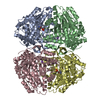
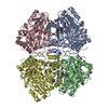
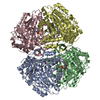
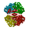
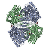
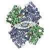
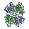
| Title | TRYPTOPHANASE FROM PROTEUS VULGARIS | ||||||
 Components Components | TRYPTOPHANASE | ||||||
 Keywords Keywords | TRYPTOPHAN BIOSYNTHESIS / TRYPTOPHAN INDOLE-LYASE / PYRIDOXAL 5'-PHOSPHATE / MONOVALENT CATION BINDING SITE | ||||||
| Function / homology |  Function and homology information Function and homology information | ||||||
| Biological species |  Proteus vulgaris (bacteria) Proteus vulgaris (bacteria) | ||||||
| Method |  X-RAY DIFFRACTION / SYNCHROTRON / MOLECULAR REPLACEMENT / Resolution: 2.1 Å X-RAY DIFFRACTION / SYNCHROTRON / MOLECULAR REPLACEMENT / Resolution: 2.1 Å | ||||||
 Authors Authors | Isupov, M.N. / Antson, A.A. / Dodson, E.J. / Dodson, G.G. / Dementieva, I.S. / Zakomirdina, L.N. / Wilson, K.S. / Dauter, Z. / Lebedev, A.A. / Harutyunyan, E.H. | ||||||
 Citation Citation | Journal: J.Mol.Biol. / Year: 1998 Title: Crystal structure of tryptophanase. Authors: Isupov, M.N. / Antson, A.A. / Dodson, E.J. / Dodson, G.G. / Dementieva, I.S. / Zakomirdina, L.N. / Wilson, K.S. / Dauter, Z. / Lebedev, A.A. / Harutyunyan, E.H. #1: Journal: Biochemistry of Vitamin B6 and Pqq / Year: 1994Title: X-Ray Study of Tryptophanase at 2.1 Angstrom Resolution Authors: Isupov, M. / Dementieva, I. / Zakomirdina, L. / Wilson, K.S. / Dauter, Z. / Antson, A.A. / Dodson, G.G. / Harutyunyan, E.H. #2: Journal: J.Mol.Biol. / Year: 1994Title: Crystallization and Preliminary X-Ray Investigation of Holotryptophanases from Escherichia Coli and Proteus Vulgaris Authors: Dementieva, I.S. / Zakomirdina, L.N. / Sinitzina, N.I. / Antson, A.A. / Wilson, K.S. / Isupov, M.N. / Lebedev, A.A. / Harutyunyan, E.H. | ||||||
| History |
|
- Structure visualization
| Structure viewer | Molecule: MolmilJmol/JSmol |
|---|
- Downloads & links
-Download
| PDBx/mmCIF format | 1ax4.cif.gz | 386.1 KB | Display | PDBx/mmCIF format |
|---|---|---|---|---|
| PDB format | pdb1ax4.ent.gz | 318.2 KB | Display | PDB format |
| PDBx/mmJSON format | 1ax4.json.gz | Tree view | PDBx/mmJSON format | |
| Others |  Other downloads Other downloads |
-Validation report
| Arichive directory | https://data.pdbj.org/pub/pdb/validation_reports/ax/1ax4ftp://data.pdbj.org/pub/pdb/validation_reports/ax/1ax4 | HTTPS FTP |
|---|
-Related structure data
| Related structure data |  1tplS S: Starting model for refinement |
|---|---|
| Similar structure data |


-Links
 PDBj
PDBj- Assembly
| Deposited unit | 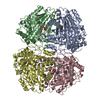
| ||||||||||||||||
|---|---|---|---|---|---|---|---|---|---|---|---|---|---|---|---|---|---|
| 1 |
| ||||||||||||||||
| Unit cell |
| ||||||||||||||||
| Noncrystallographic symmetry (NCS) | NCS oper:
|
-Components
| #1: Protein | Mass: 52787.102 Da / Num. of mol.: 4 Source method: isolated from a genetically manipulated source Details: PYRIDOXAL 5'-DEPENDENT ENZYME, REQUIRES MONOVALENT CATIONS FOR ACTIVITY, DEGRADES L-TRYPTOPHAN TO INDOLE, PYRUVATE AND AMMONIA Source: (gene. exp.) Proteus vulgaris (bacteria) / Production host: #2: Chemical | ChemComp-K /   Mass: 39.098 Da / Num. of mol.: 4 / Source method: obtained synthetically / Formula: K Mass: 39.098 Da / Num. of mol.: 4 / Source method: obtained synthetically / Formula: K#3: Water | ChemComp-HOH / |  Mass: 18.015 Da / Num. of mol.: 940 / Source method: isolated from a natural source / Formula: H2O Mass: 18.015 Da / Num. of mol.: 940 / Source method: isolated from a natural source / Formula: H2O |
|---|
-Experimental details
-Experiment
| Experiment | Method: X-RAY DIFFRACTION / Number of used crystals: 1 |
|---|
- Sample preparation
| Crystal | Density Matthews: 2.49 Å3/Da / Density % sol: 51 % | ||||||||||||||||||||||||||||||||||||
|---|---|---|---|---|---|---|---|---|---|---|---|---|---|---|---|---|---|---|---|---|---|---|---|---|---|---|---|---|---|---|---|---|---|---|---|---|---|
| Crystal grow | pH: 7.8 Details: 27 % PEG 4000, 0.1 M POTASSIUM PHOSPHATE BUFFER PH 7.8, 0.25 MM PYRIDOXAL 5'-PHOSPHATE, 5MM DTT, 0.1 M CSCL. | ||||||||||||||||||||||||||||||||||||
| Crystal grow | *PLUS Method: vapor diffusion, hanging drop | ||||||||||||||||||||||||||||||||||||
| Components of the solutions | *PLUS
|
-Data collection
| Diffraction | Mean temperature: 293 K |
|---|---|
| Diffraction source | Source: SYNCHROTRON / Site: EMBL/DESY, HAMBURG  / Beamline: BW7B / Wavelength: 0.95 / Beamline: BW7B / Wavelength: 0.95 |
| Detector | Type: MARRESEARCH / Detector: IMAGE PLATE / Date: Jan 1, 1994 |
| Radiation | Monochromatic (M) / Laue (L): M / Scattering type: x-ray |
| Radiation wavelength | Wavelength: 0.95 Å / Relative weight: 1 |
| Reflection | Resolution: 2.1→18 Å / Num. obs: 397510 / % possible obs: 97.6 % / Redundancy: 3.33 % / Rmerge(I) obs: 0.098 / Rsym value: 0.098 / Net I/σ(I): 11 |
| Reflection shell | Resolution: 2.1→2.17 Å / Redundancy: 2.49 % / Rmerge(I) obs: 0.273 / Mean I/σ(I) obs: 3.2 / Rsym value: 0.273 / % possible all: 97.3 |
| Reflection | *PLUS Num. obs: 117863 / Num. measured all: 397510 |
| Reflection shell | *PLUS % possible obs: 97.3 % |
- Processing
| Software |
| ||||||||||||||||||||||||||||||||||||||||||||||||
|---|---|---|---|---|---|---|---|---|---|---|---|---|---|---|---|---|---|---|---|---|---|---|---|---|---|---|---|---|---|---|---|---|---|---|---|---|---|---|---|---|---|---|---|---|---|---|---|---|---|
| Refinement | Method to determine structure: MOLECULAR REPLACEMENT Starting model: TYROSINE PHENOL LYASE, PDB ENTRY 1TPL Resolution: 2.1→18 Å / Cross valid method: FREE R
| ||||||||||||||||||||||||||||||||||||||||||||||||
| Refinement step | Cycle: LAST / Resolution: 2.1→18 Å
| ||||||||||||||||||||||||||||||||||||||||||||||||
| Software | *PLUS Name: REFMAC / Classification: refinement | ||||||||||||||||||||||||||||||||||||||||||||||||
| Refinement | *PLUS Num. reflection all: 117863 / Rfactor obs: 0.187 | ||||||||||||||||||||||||||||||||||||||||||||||||
| Solvent computation | *PLUS | ||||||||||||||||||||||||||||||||||||||||||||||||
| Displacement parameters | *PLUS | ||||||||||||||||||||||||||||||||||||||||||||||||
| Refine LS restraints | *PLUS
|
