+ Open data
- Basic information
| Entry | Database: PDB / ID: 1aop | ||||||||||||
|---|---|---|---|---|---|---|---|---|---|---|---|---|---|
| Title | SULFITE REDUCTASE STRUCTURE AT 1.6 ANGSTROM RESOLUTION | ||||||||||||
 Components Components | SULFITE REDUCTASE HEMOPROTEIN | ||||||||||||
 Keywords Keywords | OXIDOREDUCTASE / SIROHEME / [4FE-4S] / SNIRR / SIX-ELECTRON REDUCTION / PHOSPHATE COMPLEX | ||||||||||||
| Function / homology |  Function and homology information Function and homology informationassimilatory sulfite reductase (NADPH) / sulfite reductase (NADPH) activity / sulfite reductase complex (NADPH) / sulfate assimilation / hydrogen sulfide biosynthetic process / cysteine biosynthetic process / NADP binding / 4 iron, 4 sulfur cluster binding / heme binding / metal ion binding Similarity search - Function | ||||||||||||
| Biological species |  | ||||||||||||
| Method |  X-RAY DIFFRACTION / X-RAY DIFFRACTION /  SYNCHROTRON / MAD/MIR / Resolution: 1.6 Å SYNCHROTRON / MAD/MIR / Resolution: 1.6 Å | ||||||||||||
 Authors Authors | Crane, B.R. / Getzoff, E.D. | ||||||||||||
 Citation Citation |  Journal: Science / Year: 1995 Journal: Science / Year: 1995Title: Sulfite reductase structure at 1.6 A: evolution and catalysis for reduction of inorganic anions. Authors: Crane, B.R. / Siegel, L.M. / Getzoff, E.D. #1:  Journal: Biochemistry / Year: 1997 Journal: Biochemistry / Year: 1997Title: Structures of the Siroheme-and Fe4S4-Containing Active Center of Sulfite Reductase in Different States of Oxidation: Heme Activation Via Reduction-Gated Exogenous Ligand Exchange Authors: Crane, B.R. / Siegel, L.M. / Getzoff, E.D. #2:  Journal: Acta Crystallogr.,Sect.D / Year: 1997 Journal: Acta Crystallogr.,Sect.D / Year: 1997Title: Multiwavelength Anomalous Diffraction of Sulfite Reductase Hemoprotein: Making the Most of MAD Data Authors: Crane, B.R. / Bellamy, H. / Getzoff, E.D. #3:  Journal: Acta Crystallogr.,Sect.D / Year: 1997 Journal: Acta Crystallogr.,Sect.D / Year: 1997Title: Determining Phases and Anomalous-Scattering Models from the Multiwavelength Anomalous Diffraction of Native Protein Metal Clusters. Improved MAD Phase Error Estimates and Anomalous-Scatterer Positions Authors: Crane, B.R. / Getzoff, E.D. #4:  Journal: J.Biol.Chem. / Year: 1989 Journal: J.Biol.Chem. / Year: 1989Title: Characterization of the Cysjih Regions of Salmonella Typhimurium and Escherichia Coli B. DNA Sequences of Cysi and Cysh and a Model for the Siroheme-Fe4S4 Active Center of Sulfite Reductase ...Title: Characterization of the Cysjih Regions of Salmonella Typhimurium and Escherichia Coli B. DNA Sequences of Cysi and Cysh and a Model for the Siroheme-Fe4S4 Active Center of Sulfite Reductase Hemoprotein Based on Amino Acid Homology with Spinach Nitrite Reductase Authors: Ostrowski, J. / Wu, J.Y. / Rueger, D.C. / Miller, B.E. / Siegel, L.M. / Kredich, N.M. #5:  Journal: J.Biol.Chem. / Year: 1986 Journal: J.Biol.Chem. / Year: 1986Title: The Heme and Fe4S4 Cluster in the Crystallographic Structure of Escherichia Coli Sulfite Reductase Authors: Mcree, D.E. / Richardson, D.C. / Richardson, J.S. / Siegel, L.M. | ||||||||||||
| History |
|
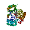
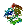
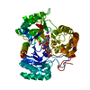
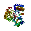
- Structure visualization
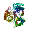
| Structure viewer | Molecule:  Molmil Molmil Jmol/JSmol Jmol/JSmol |
|---|
- Downloads & links
- Download
| PDBx/mmCIF format |  1aop.cif.gz 1aop.cif.gz | 121.9 KB | Display |  PDBx/mmCIF format PDBx/mmCIF format |
|---|---|---|---|---|
| PDB format |  pdb1aop.ent.gz pdb1aop.ent.gz | 89.9 KB | Display |  PDB format PDB format |
| PDBx/mmJSON format |  1aop.json.gz 1aop.json.gz | Tree view |  PDBx/mmJSON format PDBx/mmJSON format | |
| Others |  Other downloads Other downloads |
-Validation report
| Summary document |  1aop_validation.pdf.gz 1aop_validation.pdf.gz | 526.1 KB | Display |  wwPDB validaton report wwPDB validaton report |
|---|---|---|---|---|
| Full document |  1aop_full_validation.pdf.gz 1aop_full_validation.pdf.gz | 531.9 KB | Display | |
| Data in XML |  1aop_validation.xml.gz 1aop_validation.xml.gz | 11.8 KB | Display | |
| Data in CIF |  1aop_validation.cif.gz 1aop_validation.cif.gz | 20.3 KB | Display | |
| Arichive directory |  https://data.pdbj.org/pub/pdb/validation_reports/ao/1aop https://data.pdbj.org/pub/pdb/validation_reports/ao/1aop ftp://data.pdbj.org/pub/pdb/validation_reports/ao/1aop ftp://data.pdbj.org/pub/pdb/validation_reports/ao/1aop | HTTPS FTP |
-Related structure data
| Similar structure data |
|---|
- Links
Links
- Assembly
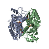
| Deposited unit | 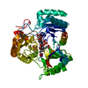
| ||||||||
|---|---|---|---|---|---|---|---|---|---|
| 1 |
| ||||||||
| Unit cell |
|
- Components
-Protein , 1 types, 1 molecules A
| #1: Protein | Mass: 55747.715 Da / Num. of mol.: 1 Source method: isolated from a genetically manipulated source Details: OXIDIZED, SIROHEME FE(III), [4FE-4S], +2 / Source: (gene. exp.)  Description: PBR322 DERIVATIVE CONTAINING ESCHERICHIA COLI CYSIJ AND S. TYPHIMURIUM CYSG UNDER CONTROL OF CYSJIH PROMOTER EXPRESSED IN A S. TYPHIMURIUM CYSI AUXOTROPH Gene: CYSIJ / Plasmid: PJYW613 / Production host:  References: UniProt: P17846, assimilatory sulfite reductase (NADPH) |
|---|
-Non-polymers , 5 types, 490 molecules 


| #2: Chemical | ChemComp-PO4 / |
|---|---|
| #3: Chemical | ChemComp-K / |
| #4: Chemical | ChemComp-SF4 / |
| #5: Chemical | ChemComp-SRM / |
| #6: Water | ChemComp-HOH / |
-Details
| Nonpolymer details | OXIDIZED, SIROHEME FE(III), [4FE-4S], +2. |
|---|
-Experimental details
-Experiment
| Experiment | Method:  X-RAY DIFFRACTION / Number of used crystals: 2 X-RAY DIFFRACTION / Number of used crystals: 2 |
|---|
- Sample preparation
| Crystal | Density Matthews: 2.13 Å3/Da / Density % sol: 40 % | |||||||||||||||||||||||||
|---|---|---|---|---|---|---|---|---|---|---|---|---|---|---|---|---|---|---|---|---|---|---|---|---|---|---|
| Crystal grow | pH: 7.7 / Details: pH 7.7 | |||||||||||||||||||||||||
| Crystal grow | *PLUS Method: vapor diffusion | |||||||||||||||||||||||||
| Components of the solutions | *PLUS
|
-Data collection
| Diffraction | Mean temperature: 277 K |
|---|---|
| Diffraction source | Source:  SYNCHROTRON / Site: SYNCHROTRON / Site:  SSRL SSRL  / Beamline: BL7-1 / Wavelength: 1.08 / Beamline: BL7-1 / Wavelength: 1.08 |
| Detector | Type: MARRESEARCH / Detector: IMAGE PLATE / Date: Feb 11, 1994 |
| Radiation | Monochromatic (M) / Laue (L): M / Scattering type: x-ray |
| Radiation wavelength | Wavelength: 1.08 Å / Relative weight: 1 |
| Reflection | Resolution: 1.6→30 Å / Num. obs: 61005 / % possible obs: 96.6 % / Observed criterion σ(I): 0 / Redundancy: 6 % / Rsym value: 0.099 / Net I/σ(I): 28.7 |
| Reflection shell | Resolution: 1.6→1.76 Å / Mean I/σ(I) obs: 7.5 / Rsym value: 0.278 / % possible all: 93.5 |
| Reflection | *PLUS Rmerge(I) obs: 0.099 |
| Reflection shell | *PLUS % possible obs: 93.5 % / Rmerge(I) obs: 0.278 |
- Processing
| Software |
| ||||||||||||||||||||||||||||||||||||||||||||||||||||||||||||
|---|---|---|---|---|---|---|---|---|---|---|---|---|---|---|---|---|---|---|---|---|---|---|---|---|---|---|---|---|---|---|---|---|---|---|---|---|---|---|---|---|---|---|---|---|---|---|---|---|---|---|---|---|---|---|---|---|---|---|---|---|---|
| Refinement | Method to determine structure: MAD/MIR / Resolution: 1.6→10 Å / σ(F): 0
| ||||||||||||||||||||||||||||||||||||||||||||||||||||||||||||
| Displacement parameters | Biso mean: 19.8 Å2
| ||||||||||||||||||||||||||||||||||||||||||||||||||||||||||||
| Refine analyze | Luzzati sigma a obs: 0.14 Å | ||||||||||||||||||||||||||||||||||||||||||||||||||||||||||||
| Refinement step | Cycle: LAST / Resolution: 1.6→10 Å
| ||||||||||||||||||||||||||||||||||||||||||||||||||||||||||||
| Refine LS restraints |
| ||||||||||||||||||||||||||||||||||||||||||||||||||||||||||||
| LS refinement shell | Resolution: 1.6→1.76 Å
| ||||||||||||||||||||||||||||||||||||||||||||||||||||||||||||
| Xplor file |
| ||||||||||||||||||||||||||||||||||||||||||||||||||||||||||||
| Software | *PLUS Name:  X-PLOR / Classification: refinement X-PLOR / Classification: refinement | ||||||||||||||||||||||||||||||||||||||||||||||||||||||||||||
| Refinement | *PLUS | ||||||||||||||||||||||||||||||||||||||||||||||||||||||||||||
| Solvent computation | *PLUS | ||||||||||||||||||||||||||||||||||||||||||||||||||||||||||||
| Displacement parameters | *PLUS | ||||||||||||||||||||||||||||||||||||||||||||||||||||||||||||
| LS refinement shell | *PLUS Rfactor obs: 0.278 |
 Movie
Movie Controller



 PDBj
PDBj


