 Movie
Movie Controller

[English] 日本語
 Yorodumi
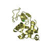
- PDB-1alc: REFINED STRUCTURE OF BABOON ALPHA-LACTALBUMIN AT 1.7 ANGSTROMS RE... -
+ Open data

- Basic information
| Entry | Database: PDB / ID: 1alc | ||||||
|---|---|---|---|---|---|---|---|
| Title | REFINED STRUCTURE OF BABOON ALPHA-LACTALBUMIN AT 1.7 ANGSTROMS RESOLUTION. COMPARISON WITH C-TYPE LYSOZYME | ||||||
 Components Components | ALPHA-LACTALBUMIN | ||||||
 Keywords Keywords | CALCIUM BINDING PROTEIN | ||||||
| Function / homology |  Function and homology information Function and homology informationlactose synthase activity / lactose biosynthetic process / lysozyme activity / defense response to Gram-negative bacterium / defense response to Gram-positive bacterium / calcium ion binding / extracellular region Similarity search - Function | ||||||
| Biological species |  | ||||||
| Method |  X-RAY DIFFRACTION / Resolution: 1.7 Å X-RAY DIFFRACTION / Resolution: 1.7 Å | ||||||
 Authors Authors | Acharya, K.R. / Stuart, D.I. / Phillips, D.C. | ||||||
 Citation Citation | Journal: J.Mol.Biol. / Year: 1989 Title: Refined structure of baboon alpha-lactalbumin at 1.7 A resolution. Comparison with C-type lysozyme. Authors: Acharya, K.R. / Stuart, D.I. / Walker, N.P. / Lewis, M. / Phillips, D.C. #1: Journal: Biochem.J. / Year: 1987Title: Crystallographic Analysis of the Three-Dimensional Structure of Baboon Alpha-Lactalbumin at Low Resolution. Homology with Lysozyme Authors: Smith, S.G. / Lewis, M. / Aschaffenburg, R. / Fenna, R.E. / Wilson, I.A. / Sundaralingam, M. / Stuart, D.I. / Phillips, D.C. #2: Journal: Nature / Year: 1986Title: Alpha-Lactalbumin Possesses a Novel Calcium Binding Loop Authors: Stuart, D.I. / Acharya, K.R. / Walker, N.P.C. / Smith, S.G. / Lewis, M. / Phillips, D.C. #3: Journal: J.Mol.Biol. / Year: 1979Title: Crystallography of Alpha-Lactalbumin. III. Crystals of Baboon Milk Alpha-Lactalbumin Authors: Aschaffenburg, R. / Fenna, R.E. / Phillips, D.C. / Smith, S.G. / Buss, D.H. / Jeness, R. / Thompson, M.P. | ||||||
| History |
|
- Structure visualization
| Structure viewer | Molecule: MolmilJmol/JSmol |
|---|
- Downloads & links
-Download
| PDBx/mmCIF format | 1alc.cif.gz | 41.9 KB | Display | PDBx/mmCIF format |
|---|---|---|---|---|
| PDB format | pdb1alc.ent.gz | 28.7 KB | Display | PDB format |
| PDBx/mmJSON format | 1alc.json.gz | Tree view | PDBx/mmJSON format | |
| Others |  Other downloads Other downloads |
-Validation report
| Arichive directory | https://data.pdbj.org/pub/pdb/validation_reports/al/1alcftp://data.pdbj.org/pub/pdb/validation_reports/al/1alc | HTTPS FTP |
|---|
-Related structure data
| Similar structure data |
|---|


-Links
 PDBj
PDBj- Assembly
| Deposited unit | 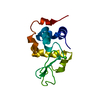
| ||||||||
|---|---|---|---|---|---|---|---|---|---|
| 1 |
| ||||||||
| Unit cell |
|
-Components
| #1: Protein | Mass: 14189.021 Da / Num. of mol.: 1 Source method: isolated from a genetically manipulated source Source: (gene. exp.) |
|---|---|
| #2: Chemical | ChemComp-CA /   Mass: 40.078 Da / Num. of mol.: 1 / Source method: obtained synthetically / Formula: Ca Mass: 40.078 Da / Num. of mol.: 1 / Source method: obtained synthetically / Formula: Ca |
| #3: Water | ChemComp-HOH /  Mass: 18.015 Da / Num. of mol.: 150 / Source method: isolated from a natural source / Formula: H2O Mass: 18.015 Da / Num. of mol.: 150 / Source method: isolated from a natural source / Formula: H2O |
| Has protein modification | Y |
-Experimental details
-Experiment
| Experiment | Method: X-RAY DIFFRACTION |
|---|
- Sample preparation
| Crystal | Density Matthews: 1.99 Å3/Da / Density % sol: 38.26 % | |||||||||||||||
|---|---|---|---|---|---|---|---|---|---|---|---|---|---|---|---|---|
| Crystal grow | *PLUS pH: 6.8 / Method: microdialysis | |||||||||||||||
| Components of the solutions | *PLUS
|
-Data collection
| Radiation | Scattering type: x-ray |
|---|---|
| Radiation wavelength | Relative weight: 1 |
| Reflection | *PLUS Highest resolution: 1.7 Å / Lowest resolution: 9999 Å / Num. all: 13025 / Num. obs: 12400 / Observed criterion σ(F): 2 / Num. measured all: 10275 |
| Reflection shell | *PLUS Highest resolution: 1.7 Å |
- Processing
| Software | Name: PROLSQ / Classification: refinement | ||||||||||||||||||||||||||||||||||||||||||||||||||||||||||||||||||||||||||||||||||||
|---|---|---|---|---|---|---|---|---|---|---|---|---|---|---|---|---|---|---|---|---|---|---|---|---|---|---|---|---|---|---|---|---|---|---|---|---|---|---|---|---|---|---|---|---|---|---|---|---|---|---|---|---|---|---|---|---|---|---|---|---|---|---|---|---|---|---|---|---|---|---|---|---|---|---|---|---|---|---|---|---|---|---|---|---|---|
| Refinement | Highest resolution: 1.7 Å /
| ||||||||||||||||||||||||||||||||||||||||||||||||||||||||||||||||||||||||||||||||||||
| Refinement step | Cycle: LAST / Highest resolution: 1.7 Å
| ||||||||||||||||||||||||||||||||||||||||||||||||||||||||||||||||||||||||||||||||||||
| Refine LS restraints |
| ||||||||||||||||||||||||||||||||||||||||||||||||||||||||||||||||||||||||||||||||||||
| Refinement | *PLUS Rfactor obs: 0.22 / Highest resolution: 1.7 Å / Lowest resolution: 9999 Å / Num. reflection obs: 10275 | ||||||||||||||||||||||||||||||||||||||||||||||||||||||||||||||||||||||||||||||||||||
| Solvent computation | *PLUS | ||||||||||||||||||||||||||||||||||||||||||||||||||||||||||||||||||||||||||||||||||||
| Displacement parameters | *PLUS |
