 Movie
Movie Controller


+ Open data

- Basic information
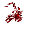
| Entry | Database: PDB / ID: 1a6r | ||||||
|---|---|---|---|---|---|---|---|
| Title | GAL6 (YEAST BLEOMYCIN HYDROLASE) MUTANT C73A | ||||||
 Components Components | GAL6 | ||||||
 Keywords Keywords | HYDROLASE / BLEOMYCIN HYDROLASE / PEPTIDASE / PROTEASE / DNA-BINDING PROTEIN / SELF-COMPARTMENTALIZING PROTEASE | ||||||
| Function / homology |  Function and homology information Function and homology informationbleomycin hydrolase / cysteine-type aminopeptidase activity / L-homocysteine catabolic process / Antigen processing: Ubiquitination & Proteasome degradation / aminopeptidase activity / cysteine-type peptidase activity / response to toxic substance / single-stranded DNA binding / double-stranded DNA binding / RNA polymerase II cis-regulatory region sequence-specific DNA binding ...bleomycin hydrolase / cysteine-type aminopeptidase activity / L-homocysteine catabolic process / Antigen processing: Ubiquitination & Proteasome degradation / aminopeptidase activity / cysteine-type peptidase activity / response to toxic substance / single-stranded DNA binding / double-stranded DNA binding / RNA polymerase II cis-regulatory region sequence-specific DNA binding / cysteine-type endopeptidase activity / response to antibiotic / mRNA binding / negative regulation of transcription by RNA polymerase II / mitochondrion / proteolysis / cytoplasm Similarity search - Function | ||||||
| Biological species |  | ||||||
| Method |  X-RAY DIFFRACTION / SYNCHROTRON / MOLECULAR REPLACEMENT / Resolution: 2.05 Å X-RAY DIFFRACTION / SYNCHROTRON / MOLECULAR REPLACEMENT / Resolution: 2.05 Å | ||||||
 Authors Authors | Joshua-Tor, L. / Zheng, W. / Johnston, S.A. | ||||||
 Citation Citation | Journal: Cell(Cambridge,Mass.) / Year: 1998 Title: The unusual active site of Gal6/bleomycin hydrolase can act as a carboxypeptidase, aminopeptidase, and peptide ligase. Authors: Zheng, W. / Johnston, S.A. / Joshua-Tor, L. #1: Journal: Science / Year: 1995Title: Crystal Structure of a Conserved Protease that Binds DNA: The Bleomycin Hydrolase, Gal6 Authors: Joshua-Tor, L. / Xu, H.E. / Johnston, S.A. / Rees, D.C. | ||||||
| History |
|
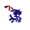
- Structure visualization
| Structure viewer | Molecule: MolmilJmol/JSmol |
|---|
- Downloads & links
-Download
| PDBx/mmCIF format | 1a6r.cif.gz | 114.1 KB | Display | PDBx/mmCIF format |
|---|---|---|---|---|
| PDB format | pdb1a6r.ent.gz | 86.8 KB | Display | PDB format |
| PDBx/mmJSON format | 1a6r.json.gz | Tree view | PDBx/mmJSON format | |
| Others |  Other downloads Other downloads |
-Validation report
| Arichive directory | https://data.pdbj.org/pub/pdb/validation_reports/a6/1a6rftp://data.pdbj.org/pub/pdb/validation_reports/a6/1a6r | HTTPS FTP |
|---|
-Related structure data
| Related structure data |  3gcbC  1gcbS S: Starting model for refinement C: citing same article ( |
|---|---|
| Similar structure data |


-Links
 PDBj
PDBj


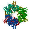
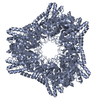
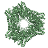
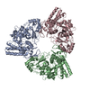
- Assembly
| Deposited unit | 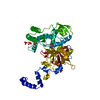
| ||||||||
|---|---|---|---|---|---|---|---|---|---|
| 1 | x 6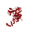
| ||||||||
| Unit cell |
|
-Components
| #1: Protein | Mass: 54053.449 Da / Num. of mol.: 1 / Mutation: HIS6-TEV-TAG, S2A, C73A Source method: isolated from a genetically manipulated source Source: (gene. exp.) Cell line: BL21 / Gene: GAL6C73A / Plasmid: PKM260/PLYSS / Species (production host): Escherichia coli / Gene (production host): GAL6C73A / Production host:  References: UniProt: Q01532, Hydrolases; Acting on peptide bonds (peptidases); Cysteine endopeptidases | ||
|---|---|---|---|
| #2: Chemical | ChemComp-SO4 /   Mass: 96.063 Da / Num. of mol.: 6 / Source method: obtained synthetically / Formula: SO4 Mass: 96.063 Da / Num. of mol.: 6 / Source method: obtained synthetically / Formula: SO4#3: Water | ChemComp-HOH / |  Mass: 18.015 Da / Num. of mol.: 370 / Source method: isolated from a natural source / Formula: H2O Mass: 18.015 Da / Num. of mol.: 370 / Source method: isolated from a natural source / Formula: H2O |
-Experimental details
-Experiment
| Experiment | Method: X-RAY DIFFRACTION / Number of used crystals: 1 |
|---|
- Sample preparation
| Crystal | Density Matthews: 2.73 Å3/Da / Density % sol: 55 % | |||||||||||||||||||||||||||||||||||
|---|---|---|---|---|---|---|---|---|---|---|---|---|---|---|---|---|---|---|---|---|---|---|---|---|---|---|---|---|---|---|---|---|---|---|---|---|
| Crystal grow | pH: 9 Details: PROTEIN WAS CRYSTALLIZED FROM AMMONIUM SULFATE AND PEG 4K., pH 9.0 | |||||||||||||||||||||||||||||||||||
| Crystal | *PLUS | |||||||||||||||||||||||||||||||||||
| Crystal grow | *PLUS pH: 8.5 / Method: vapor diffusion, sitting drop | |||||||||||||||||||||||||||||||||||
| Components of the solutions | *PLUS
|
-Data collection
| Diffraction | Mean temperature: 90 K |
|---|---|
| Diffraction source | Source: SYNCHROTRON / Site: NSLS  / Beamline: X12B / Wavelength: 1.08 / Beamline: X12B / Wavelength: 1.08 |
| Detector | Type: MAR scanner 300 mm plate / Detector: IMAGE PLATE / Date: Apr 1, 1996 |
| Radiation | Monochromatic (M) / Laue (L): M / Scattering type: x-ray |
| Radiation wavelength | Wavelength: 1.08 Å / Relative weight: 1 |
| Reflection | Resolution: 2.05→50 Å / Num. obs: 37272 / % possible obs: 97.3 % / Observed criterion σ(I): 0 / Redundancy: 9.8 % / Biso Wilson estimate: 13.8 Å2 / Rmerge(I) obs: 0.077 / Rsym value: 0.077 / Net I/σ(I): 58 |
| Reflection shell | Resolution: 2.05→2.12 Å / Rmerge(I) obs: 0.164 / Mean I/σ(I) obs: 31 / Rsym value: 0.164 / % possible all: 99.2 |
| Reflection | *PLUS Num. measured all: 367565 |
| Reflection shell | *PLUS % possible obs: 99.2 % |
- Processing
| Software |
| ||||||||||||||||||||||||||||||||||||||||||||||||||||||||||||||||||||||||||||||||
|---|---|---|---|---|---|---|---|---|---|---|---|---|---|---|---|---|---|---|---|---|---|---|---|---|---|---|---|---|---|---|---|---|---|---|---|---|---|---|---|---|---|---|---|---|---|---|---|---|---|---|---|---|---|---|---|---|---|---|---|---|---|---|---|---|---|---|---|---|---|---|---|---|---|---|---|---|---|---|---|---|---|
| Refinement | Method to determine structure: MOLECULAR REPLACEMENT Starting model: 1GCB Resolution: 2.05→8 Å / Data cutoff low absF: 0 / Isotropic thermal model: RESTRAINED / Cross valid method: THROUGHOUT / σ(F): 0 Details: 46 PROTEIN ATOMS WITH 0 OCCUPANCY BECAUSE OF DISORDERED SIDE CHAINS
| ||||||||||||||||||||||||||||||||||||||||||||||||||||||||||||||||||||||||||||||||
| Displacement parameters | Biso mean: 16.8 Å2 | ||||||||||||||||||||||||||||||||||||||||||||||||||||||||||||||||||||||||||||||||
| Refinement step | Cycle: LAST / Resolution: 2.05→8 Å
| ||||||||||||||||||||||||||||||||||||||||||||||||||||||||||||||||||||||||||||||||
| Refine LS restraints |
| ||||||||||||||||||||||||||||||||||||||||||||||||||||||||||||||||||||||||||||||||
| LS refinement shell | Resolution: 2.05→2.12 Å / Total num. of bins used: 10
| ||||||||||||||||||||||||||||||||||||||||||||||||||||||||||||||||||||||||||||||||
| Xplor file |
| ||||||||||||||||||||||||||||||||||||||||||||||||||||||||||||||||||||||||||||||||
| Software | *PLUS Name: X-PLOR / Version: 3.1 / Classification: refinement | ||||||||||||||||||||||||||||||||||||||||||||||||||||||||||||||||||||||||||||||||
| Refine LS restraints | *PLUS
|
