 Movie
Movie Controller

[English] 日本語
 Yorodumi
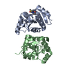
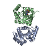
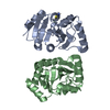
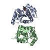
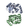
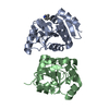
- PDB-1a4x: PYRR, THE BACILLUS SUBTILIS PYRIMIDINE BIOSYNTHETIC OPERON REPRES... -
+ Open data

- Basic information
| Entry | Database: PDB / ID: 1a4x | ||||||
|---|---|---|---|---|---|---|---|
| Title | PYRR, THE BACILLUS SUBTILIS PYRIMIDINE BIOSYNTHETIC OPERON REPRESSOR, HEXAMERIC FORM | ||||||
 Components Components | PYRIMIDINE OPERON REGULATORY PROTEIN PYRR | ||||||
 Keywords Keywords | TRANSCRIPTION REGULATION / ATTENUATION PROTEIN / RNA-BINDING PROTEIN / PYRIMIDINE BIOSYNTHESIS / TRANSFERASE / PRTASE / PHOSPHORIBOSYLTRANSFERASE / BIFUNCTIONAL ENZYME | ||||||
| Function / homology |  Function and homology information Function and homology informationuracil phosphoribosyltransferase / uracil phosphoribosyltransferase activity / DNA-templated transcription termination / RNA binding Similarity search - Function | ||||||
| Biological species |  | ||||||
| Method |  X-RAY DIFFRACTION / MOLECULAR REPLACEMENT / Resolution: 2.3 Å X-RAY DIFFRACTION / MOLECULAR REPLACEMENT / Resolution: 2.3 Å | ||||||
 Authors Authors | Tomchick, D.R. / Turner, R.J. / Switzer, R.W. / Smith, J.L. | ||||||
 Citation Citation | Journal: Structure / Year: 1998 Title: Adaptation of an enzyme to regulatory function: structure of Bacillus subtilis PyrR, a pyr RNA-binding attenuation protein and uracil phosphoribosyltransferase. Authors: Tomchick, D.R. / Turner, R.J. / Switzer, R.L. / Smith, J.L. | ||||||
| History |
|
- Structure visualization
| Structure viewer | Molecule: MolmilJmol/JSmol |
|---|
- Downloads & links
-Download
| PDBx/mmCIF format | 1a4x.cif.gz | 83.1 KB | Display | PDBx/mmCIF format |
|---|---|---|---|---|
| PDB format | pdb1a4x.ent.gz | 64.1 KB | Display | PDB format |
| PDBx/mmJSON format | 1a4x.json.gz | Tree view | PDBx/mmJSON format | |
| Others |  Other downloads Other downloads |
-Validation report
| Arichive directory | https://data.pdbj.org/pub/pdb/validation_reports/a4/1a4xftp://data.pdbj.org/pub/pdb/validation_reports/a4/1a4x | HTTPS FTP |
|---|
-Related structure data
| Related structure data |  1a3cSC S: Starting model for refinement C: citing same article ( |
|---|---|
| Similar structure data |


-Links
 PDBj
PDBj
- Assembly
| Deposited unit | 
| |||||||||
|---|---|---|---|---|---|---|---|---|---|---|
| 1 |
| |||||||||
| Unit cell |
| |||||||||
| Noncrystallographic symmetry (NCS) | NCS domain:
NCS oper: (Code: given Matrix: (-0.556303, 0.830967, -0.004548), Vector: |
-Components
| #1: Protein | Mass: 20290.279 Da / Num. of mol.: 2 Source method: isolated from a genetically manipulated source Source: (gene. exp.) #2: Chemical |   Mass: 96.063 Da / Num. of mol.: 2 / Source method: obtained synthetically / Formula: SO4 Mass: 96.063 Da / Num. of mol.: 2 / Source method: obtained synthetically / Formula: SO4#3: Water | ChemComp-HOH / |  Mass: 18.015 Da / Num. of mol.: 237 / Source method: isolated from a natural source / Formula: H2O Mass: 18.015 Da / Num. of mol.: 237 / Source method: isolated from a natural source / Formula: H2O |
|---|
-Experimental details
-Experiment
| Experiment | Method: X-RAY DIFFRACTION / Number of used crystals: 1 |
|---|
- Sample preparation
| Crystal | Density Matthews: 3.4 Å3/Da / Density % sol: 58 % | ||||||||||||||||||||||||||||||||||||||||||
|---|---|---|---|---|---|---|---|---|---|---|---|---|---|---|---|---|---|---|---|---|---|---|---|---|---|---|---|---|---|---|---|---|---|---|---|---|---|---|---|---|---|---|---|
| Crystal grow | pH: 5.1 Details: PROTEIN WAS CRYSTALLIZED FROM 14-16% PEG 6000, 300 MM AMMONIUM SULFATE, 0.2% BETA-OCTYLGLUCOSIDE, 50 MM SODIUM SUCCINATE, PH 5.1. HEXAGONAL SETTING FOR R 3 2. | ||||||||||||||||||||||||||||||||||||||||||
| Crystal | *PLUS | ||||||||||||||||||||||||||||||||||||||||||
| Crystal grow | *PLUS Temperature: 20 ℃ / pH: 7.5 / Method: vapor diffusion, hanging drop | ||||||||||||||||||||||||||||||||||||||||||
| Components of the solutions | *PLUS
|
-Data collection
| Diffraction | Mean temperature: 120 K |
|---|---|
| Diffraction source | Source: ROTATING ANODE / Type: RIGAKU RUH2R / Wavelength: 1.5418 |
| Detector | Type: RIGAKU RAXIS II / Detector: IMAGE PLATE / Date: Sep 1, 1995 / Details: MIRRORS |
| Radiation | Monochromator: NI FILTER / Monochromatic (M) / Laue (L): M / Scattering type: x-ray |
| Radiation wavelength | Wavelength: 1.5418 Å / Relative weight: 1 |
| Reflection | Resolution: 2.3→40 Å / Num. obs: 23877 / % possible obs: 98.6 % / Observed criterion σ(I): 0 / Redundancy: 3.9 % / Biso Wilson estimate: 37.8 Å2 / Rsym value: 0.05 / Net I/σ(I): 34 |
| Reflection shell | Resolution: 2.3→2.4 Å / Redundancy: 3 % / Mean I/σ(I) obs: 8 / Rsym value: 0.158 / % possible all: 97.5 |
| Reflection | *PLUS Num. measured all: 92317 / Rmerge(I) obs: 0.05 |
- Processing
| Software |
| ||||||||||||||||||||||||||||||||||||||||||||||||||||||||||||
|---|---|---|---|---|---|---|---|---|---|---|---|---|---|---|---|---|---|---|---|---|---|---|---|---|---|---|---|---|---|---|---|---|---|---|---|---|---|---|---|---|---|---|---|---|---|---|---|---|---|---|---|---|---|---|---|---|---|---|---|---|---|
| Refinement | Method to determine structure: MOLECULAR REPLACEMENT Starting model: PDB ENTRY 1A3C Resolution: 2.3→15 Å / Data cutoff high absF: 10000000 / Data cutoff low absF: 0.001 / Isotropic thermal model: RESTRAINED / Cross valid method: THROUGHOUT / σ(F): 0 Details: LYS 40, IN BOTH THE A AND B CHAINS, HAS PHI/PSI VALUES THAT ARE NORMALLY DISALLOWED. IN SOME OTHER PRTASES, THIS PEPTIDE IS TYPICALLY FOUND IN THE CIS CONFORMATION.
| ||||||||||||||||||||||||||||||||||||||||||||||||||||||||||||
| Displacement parameters | Biso mean: 35.7 Å2 | ||||||||||||||||||||||||||||||||||||||||||||||||||||||||||||
| Refinement step | Cycle: LAST / Resolution: 2.3→15 Å
| ||||||||||||||||||||||||||||||||||||||||||||||||||||||||||||
| Refine LS restraints |
| ||||||||||||||||||||||||||||||||||||||||||||||||||||||||||||
| Refine LS restraints NCS | Refine-ID: X-RAY DIFFRACTION / Rms dev Biso : 30 Å2 / Rms dev position: 1 Å / Weight Biso : 0.5
| ||||||||||||||||||||||||||||||||||||||||||||||||||||||||||||
| LS refinement shell | Resolution: 2.3→2.35 Å / Total num. of bins used: 15
| ||||||||||||||||||||||||||||||||||||||||||||||||||||||||||||
| Software | *PLUS Name: X-PLOR / Version: 3.1 / Classification: refinement | ||||||||||||||||||||||||||||||||||||||||||||||||||||||||||||
| Refine LS restraints | *PLUS
|
