National Institutes of Health/National Institute of General Medical Sciences (NIH/NIGMS)
R35GM145341
United States
National Institutes of Health/National Institute of General Medical Sciences (NIH/NIGMS)
R35GM131901
United States
National Institutes of Health/National Institute of General Medical Sciences (NIH/NIGMS)
R35GM127086
United States
Citation
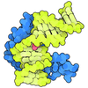
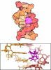
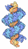
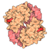
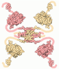
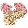
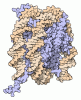
Journal: Nucleic Acids Res / Year: 2026 Title: Cryo-EM structure of a 95-base-pair double-stranded DNA minicircle at 5.3 Å resolution. Authors: Yukang Liu / Kyu-Yeon Lee / Yao He / Donggyun Kim / Hongjian Chang / Vadim Cherezov / Juli Feigon / Peter Z Qin / Abstract: Double-stranded DNA minicircles have been observed in a variety of biological settings and are also widely employed in biotechnology, therapeutic applications, and basic research. Here, we report a ...Double-stranded DNA minicircles have been observed in a variety of biological settings and are also widely employed in biotechnology, therapeutic applications, and basic research. Here, we report a cryo-EM structure of a 95-base-pair minicircle (dsMC95) at a 5.3 Å resolution. dsMC95 forms a closed ring as designed and no severe local duplex disruption is observed. The two DNA strands are fully resolved, with the major and minor grooves clearly distinguishable. Analysis reveals a nine-fold periodicity in the helical twist, which corresponds to approximately 10.56 base pairs per turn. Together with groove width analysis, the data indicate that dsMC95 maintains a B-DNA configuration. The dsMC95 ring exhibits modest in-plane ellipticity and small out-of-plane displacement, with outward-facing grooves widened and inward-facing ones compressed. The dsMC95 structure, which is the only free DNA cryo-EM structure with a resolution better than 6 Å to date, allows comparison to other structures to better understand DNA physical features such as bending. The findings advance our understanding of DNA structure under topological constraints and will inform studies of naturally occurring small circular DNA as well as the manipulation of DNA in nanotechnology applications.
History
Deposition
May 5, 2026
Deposition site: RCSB / Processing site: RCSB
Revision 1.0
Sep 30, 2026
Provider: repository / Type: Initial release
Revision 1.0
Sep 30, 2026
Data content type: EM metadata / Data content type: EM metadata / Provider: repository / Type: Initial release
Mass: 29709.711 Da / Num. of mol.: 1 / Source method: obtained synthetically Details: The deposited coordinates represent a surrogate poly(dA) sequence instead of the actual DNA sequence Source: (synth.) synthetic construct (others)
#2: DNA chain
DNA (95-MER)
Mass: 28853.314 Da / Num. of mol.: 1 / Source method: obtained synthetically Details: The deposited coordinates represent a surrogate poly(dT) sequence instead of the actual DNA sequence. Source: (synth.) synthetic construct (others)
Has protein modification
N
Sequence details
The coordinates contain an A-T only DNA model because the map does not support sequence-specific ...The coordinates contain an A-T only DNA model because the map does not support sequence-specific assignment. The actual DNA sequences are: CGATCAAGCCAGTGATAAGTGGAATGCCATGTGGTAAGATCGGTAGTCGTAGGCTCTCAA CTCGTATTCATCAACTGCATTCTGCCTACGACTAC and GTAGTCGTAGGCAGAATGCAGTTGATGAATACGAGTTGAGAGCCTACGACTACCGATCTT ACCACATGGCATTCCACTTATCACTGGCTTGATCG.
-
Experimental details
-
Experiment
Experiment
Method: ELECTRON MICROSCOPY
EM experiment
Aggregation state: PARTICLE / 3D reconstruction method: single particle reconstruction
-
Sample preparation
Component
ID
Name
Type
Entity ID
Parent-ID
Source
1
Synthetic 95-bp double-stranded DNA minicircle
COMPLEX
all
0
SYNTHETIC
2
DNA (95-MER)
COMPLEX
#1
1
SYNTHETIC
3
DNA (95-MER)
COMPLEX
#2
1
SYNTHETIC
Molecular weight
ID
Entity assembly-ID
Value (°)
Experimental value
1
1
0.0587MDa
NO
2
1
0.0293MDa
NO
3
1
0.0294MDa
NO
Source (natural)
ID
Entity assembly-ID
Organism
Ncbi tax-ID
3
2
synthetic construct (others)
32630
4
3
synthetic construct (others)
32630
Buffer solution
pH: 8 / Details: 20 mM Tris-HCl (pH 8.0) and 1 mM EDTA
Buffer component
ID
Conc.
Name
Buffer-ID
1
20mM
Tris(hydroxymethyl)aminomethanehydrochloride
1
2
1mM
Ethylenediaminetetraaceticaciddisodiumsalt
1
Specimen
Conc.: 0.7 mg/ml / Embedding applied: NO / Shadowing applied: NO / Staining applied: NO / Vitrification applied: YES
Instrument: FEI VITROBOT MARK IV / Cryogen name: ETHANE / Humidity: 100 % / Chamber temperature: 298 K
-
Electron microscopy imaging
Experimental equipment
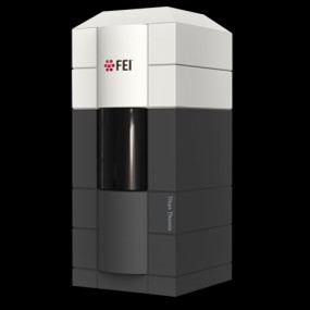
Model: Titan Krios / Image courtesy: FEI Company
Microscopy
Model: TFS KRIOS
Electron gun
Electron source: FIELD EMISSION GUN / Accelerating voltage: 300 kV / Illumination mode: FLOOD BEAM
Electron lens
Mode: BRIGHT FIELD / Nominal magnification: 165000 X / Nominal defocus max: 3000 nm / Nominal defocus min: 500 nm
Image recording
Average exposure time: 3.5 sec. / Electron dose: 50 e/Å2 / Film or detector model: GATAN K3 (6k x 4k) / Num. of grids imaged: 1 / Num. of real images: 30028
-
Processing
EM software
ID
Name
Category
1
cryoSPARC
particleselection
2
PHENIX
modelrefinement
5
cryoSPARC
CTFcorrection
12
cryoSPARC
classification
13
cryoSPARC
3Dreconstruction
CTF correction
Type: PHASE FLIPPING AND AMPLITUDE CORRECTION
Particle selection
Num. of particles selected: 1412668
Symmetry
Point symmetry: C1 (asymmetric)
3D reconstruction
Resolution: 5.27 Å / Resolution method: FSC 0.143 CUT-OFF / Num. of particles: 344629 / Num. of class averages: 1 / Symmetry type: POINT
In the structure databanks used in Yorodumi, some data are registered as the other names, "COVID-19 virus" and "2019-nCoV". Here are the details of the virus and the list of structure data.
Jan 31, 2019. EMDB accession codes are about to change! (news from PDBe EMDB page)
EMDB accession codes are about to change! (news from PDBe EMDB page)
The allocation of 4 digits for EMDB accession codes will soon come to an end. Whilst these codes will remain in use, new EMDB accession codes will include an additional digit and will expand incrementally as the available range of codes is exhausted. The current 4-digit format prefixed with “EMD-” (i.e. EMD-XXXX) will advance to a 5-digit format (i.e. EMD-XXXXX), and so on. It is currently estimated that the 4-digit codes will be depleted around Spring 2019, at which point the 5-digit format will come into force.
The EM Navigator/Yorodumi systems omit the EMD- prefix.
Related info.:Q: What is EMD? / ID/Accession-code notation in Yorodumi/EM Navigator
Yorodumi is a browser for structure data from EMDB, PDB, SASBDB, etc.
This page is also the successor to EM Navigator detail page, and also detail information page/front-end page for Omokage search.
The word "yorodu" (or yorozu) is an old Japanese word meaning "ten thousand". "mi" (miru) is to see.
Related info.:EMDB / PDB / SASBDB / Comparison of 3 databanks / Yorodumi Search / Aug 31, 2016. New EM Navigator & Yorodumi / Yorodumi Papers / Jmol/JSmol / Function and homology information / Changes in new EM Navigator and Yorodumi
 Movie
 Controller


 Open data

 Basic information
 Components
 Keywords
 Function and homology information
 Authors
 United States, 3items
 Citation
 Structure visualization
 Downloads & links
 Other downloads

 PDBj
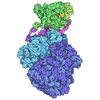
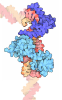
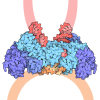
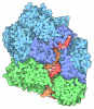

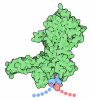
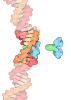
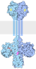
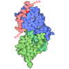
 Assembly

 Sample preparation
 Electron microscopy imaging
 FIELD EMISSION GUN / Accelerating voltage: 300 kV / Illumination mode: FLOOD BEAM
 Processing